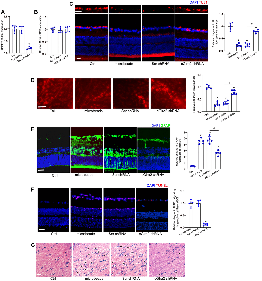
Figure 7.Silencing of cGlra2 alleviates retinal neurodegeneration in vivo. (A, B) C57BL/6 mice received intravitreous injections of cGlra2 shRNA, scrambled (Scr) shRNA, or left untreated for 14 days. qRT-PCRs were performed to examine the expression levels of cGlra2 (A, n = 5 animals) and Glra2 mRNA (B, n = 5 animals). (C) Normal retinas (Ctrl), microbeads-injected retinas, microbeads-injected retinas plus Scr shRNA injection, or microbeads-injected retinas plus cGlra2 shRNA injection were stained with TUJ1 to label RGCs. Scale bar, 20 μm; n = 5 animals. (D) Retinal whole-mounts following TUJ1 staining were observed from peripheral area. RGC survival was calculated by dividing the average number of TUJ1-positive cells in one field in the injured retina by that in control (Ctrl) retina (n = 5 animals, Scale bar: 20 μm). (E) Immunofluorescence staining with GFAP was conducted to detect retinal neurodegeneration at 2-month following microbeads injection (n = 5 animals, Scale bar: 20 μm). Green: GFAP-positive cells; Blue: DAPI. (F) TUNEL assays were performed to detect retinal apoptosis at 2-month following anterior chamber injections of microbeads (n = 5 animals, Scale bar: 50 μm). Red: TUNEL-positive cells; Blue: DAIP. (G) Degeneration of RGC axons was detected by H&E staining (n = 5 animals, Scale bar: 20 μm). Three photographs were taken at 40 × magnification for each nerve (photograph from the proximal, central, and distal portion of optic nerve). “*” in Figure 7G indicated the swellings in RGC axons. *P < 0.05 vs. Ctrl group; #P < 0.05 between the marked groups; One-way ANOVA followed by the post hoc Bonferroni test.