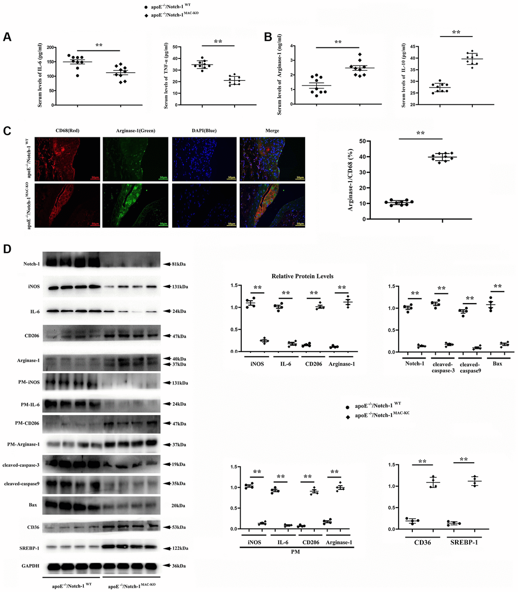
Figure 3.Notch-1MAC-KO repressed pro-inflammatory (M1) responses and stimulated anti-inflammatory (M2) effects in AS. (A, B) IL-6 and TNFα (M1 markers) were decreased in K.O. mice, whereas Arg-1 and IL-10 (M2 markers) were increased in K.O. mice. (**P < 0.01: ApoE−/−/Notch-1MAC-KO vs. ApoE−/−/Notch-1WT). (C) The aortic roots were collected, and immunofluorescent staining showed decreased staining of positive macrophages (CD68) and increased staining of Arg-1 in Notch-1MAC-KO mice. (D) Western blotting and quantitative analysis revealed down-regulated Notch-1, IL-6 (from peritoneal macrophages), iNOS (from peritoneal macrophages), cleaved-caspase-3, caspase-9 and Bax, and up-regulated CD206, Arg-1, CD36, SREBP-1, CD206 (from peritoneal macrophages) and Arg-1 (from peritoneal macrophages) in Notch-1MAC-KO mice. **P < 0.01: ApoE−/−/Notch-1MAC-KO vs. ApoE−/−/Notch-1WT.