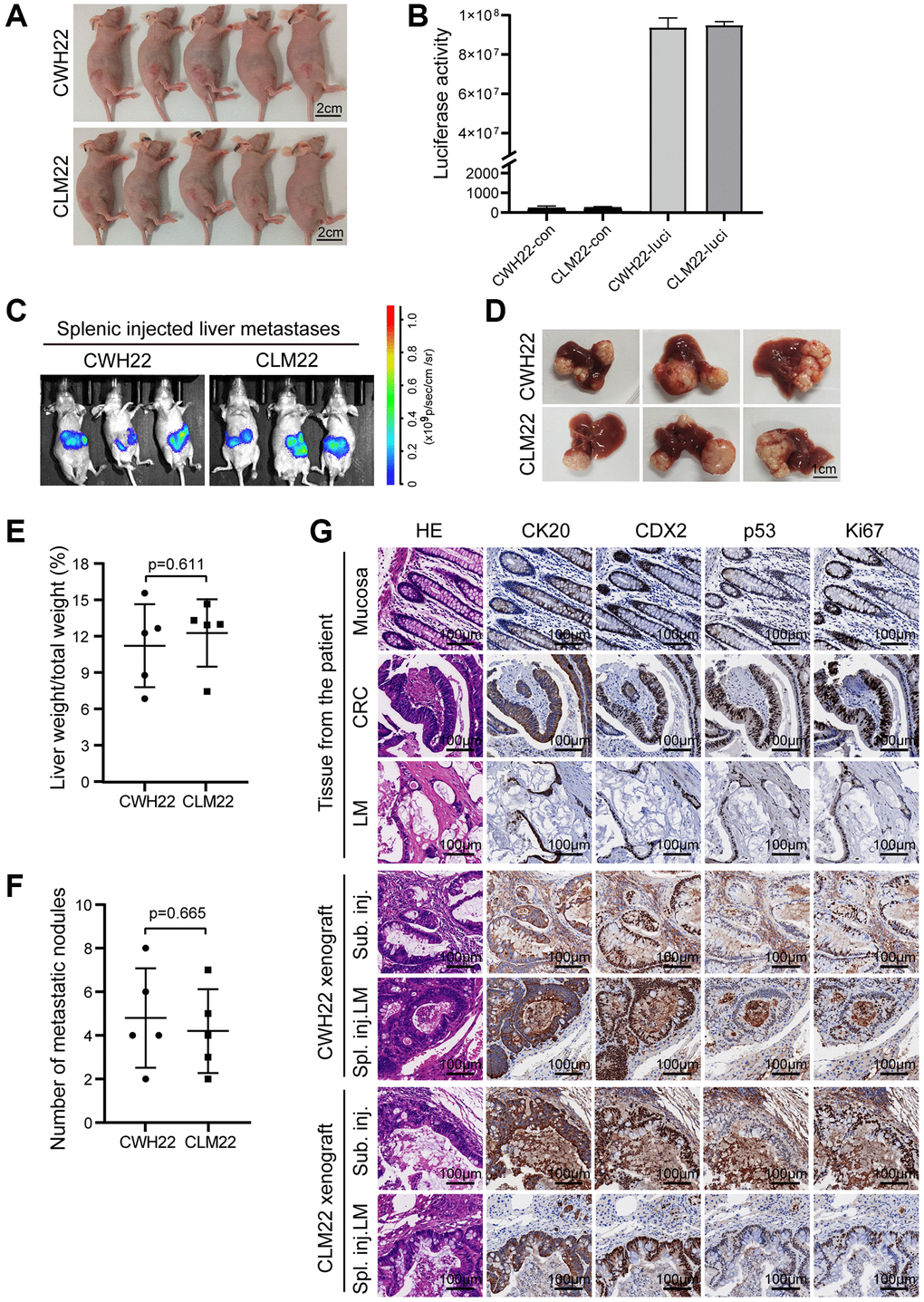
Figure 6.Tumorigenicity of CWH22 and CLM22 organoids in vivo. (A) Images of nude mice bearing CWH22 and CLM22 subcutaneous xenografts (scale bars, 2 cm). (B) Luciferase activity tests of CWH22-luci and CLM22-luci (CWH22 and CLM22 with no luciferase expression were tested as control). (C) Representative bioluminescence images of mice with liver metastases 42 days after splenic injection. (D) Representative pictures of livers with metastatic lesions (on day 45) harvested from mice receiving splenic injections of CWH22 and CLM22 organoid cells (scale bars, 1 cm). (E) Quantitative analysis of the percentage of tumor-bearing liver weight to body weight following splenic injection (n = 5; data represented as the mean ± SD). (F) Quantitative analysis of the metastatic nodule numbers per mouse following splenic injection (n = 5; data represented as the mean ± SD). (G) H&E morphology and IHC stains of tumor tissues from nude mice in A and B, as well as the normal mucosa and tumor tissues of the patient. Representative images of the expression of CK20, CDX2, Ki67, and p53 are shown (scale bars, 100 μm).