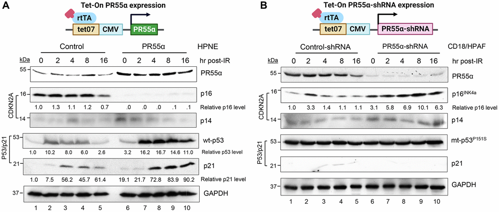
Figure 2.PR55α suppresses p16 protein expression in normal and cancer cells originating from the human exocrine pancreas. (A) Human pancreatic normal ductal (HPNE) cells were stably transduced with a retroviral vector expressing a Dox-inducible PR55α cDNA (PR55α). As a control, HPNE cells stably transduced with a relevant empty retroviral vector were included in the analysis (Control). Following the induction of ectopic PR55α expression by Dox (1 μg/ml) for 2 days, cells were exposed to 10 Gy of ionizing radiation (IR), incubated for the indicated hours, and analyzed by immunoblotting for the levels of p16, p14, p53, and p21. GAPDH in the lysates was measured as an internal control. (B) Human pancreatic ductal adenocarcinoma cells (CD18/HPAF) were stably transduced with a lentiviral vector expressing a Dox-inducible shRNA against either PR55α (PR55α-shRNA) or an irrelevant negative control (Control-shRNA). Following induction of the shRNA with Dox (2 μg/ml) for 5 days, the cells were exposed to 10 Gy IR, incubated for the indicated hours, and analyzed by immunoblotting for the levels of the indicated proteins. GAPDH in the lysates was again used as an internal control. The difference in the p16 levels between HPNE-Control and HPNE-PR55α cells, as well as between the CD18/HPAF-Control-shRNA and CD18/HPAF-PR55α-shRNA cells were determined to be statistically significant (HPNE-Control vs. HPNE-PR55α, p < 0.001; CD18/HPAF-Control-shRNA vs. CD18/HPAF-PR55α-shRNA, p = 0.004).