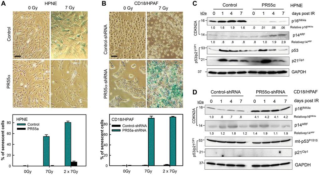
Figure 6.PR55α inhibits IR-induced cellular senescence. (A) Ectopic PR55α overexpression prevents senescence induction by IR in normal HPNE cells. HPNE/PR55α and HPNE/control cells were incubated in the presence of 1 μg/ml Dox for 2 days (to induce ectopic PR55α in the HPNE/PR55α cells) and then exposed to 7 Gy IR or left unirradiated as control (0 Gy). When the second radiation dose was applied, the interval was 24 hours between the two doses. Seven days post-IR, cells were assessed for SA-β-gal activity and photographed. Scale bar = 1 μm. The bar graph expresses the percent of senescent cells in the indicated samples and represents the mean ± S.D. of two separate experiments with each done in duplicate samples. (B) PR55α-knockdown sensitizes CD18/HPAF pancreatic cancer cells to senescence induction by IR. CD18/HPAF cells expressing Dox-inducible PR55α-shRNA or Control-shRNA were cultivated in the presence of 2 μg/ml Dox for 5 days, to allow time to silence PR55α expression, and then exposed to 7 Gy IR, or left unirradiated as a control (0 Gy). After 7 days, the cells were assessed for SA-β-gal activity and photographed. Scale bar = 1 μm. The graphs express the percent of senescent cells in the indicated samples and represent the mean ± S.D. of two separate experiments with each one in duplicate samples. (C, D) Normal HPNE and CD18/HPAF pancreatic cancer cells, with/without PR55α manipulation, were exposed to 7 Gy IR, or left unirradiated as a control (0 day). When the second radiation dose was applied, the interval was 24 hours between the two doses. The irradiated cells were incubated for the times indicated and analyzed by immunoblotting for the differences in levels of p16, p14, p53, and p21. GAPDH was used as an internal control.