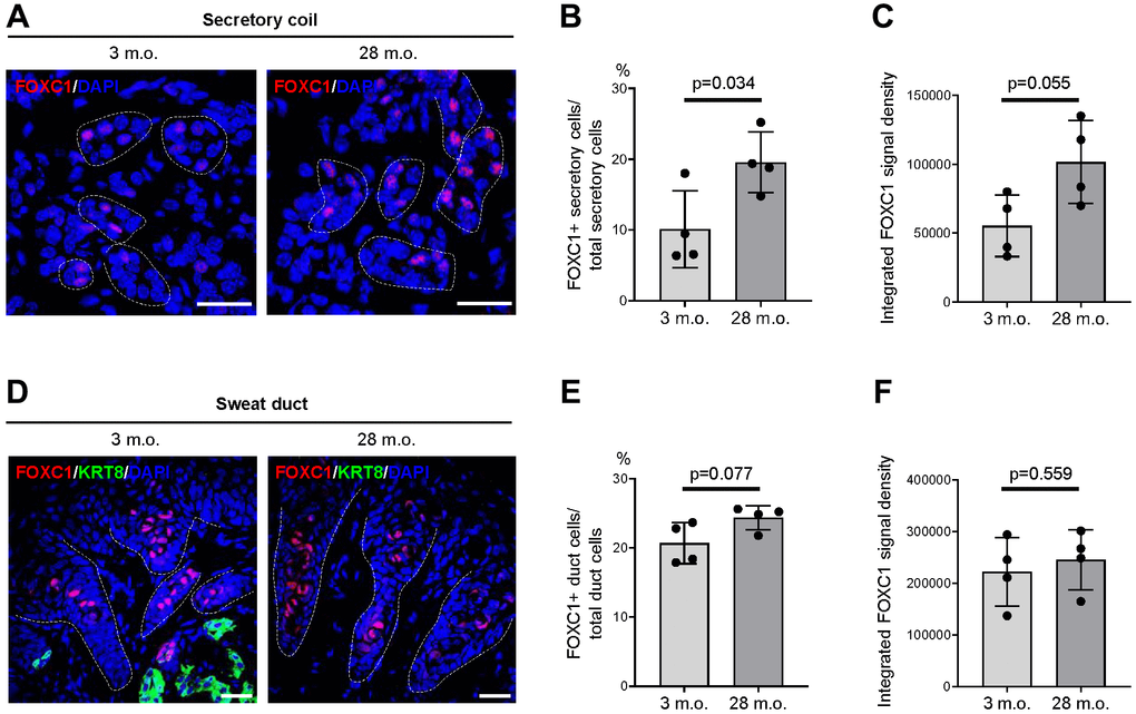
Figure 5.Expression changes of FOXC1 protein in old SWGs. (A–C) FOXC1+ secretory cells in young (3 m.o.) and old (28 m.o.) male SWGs were detected by immunofluorescence microscopy (A), and the numbers of FOXC1+ secretory cells (B), and average FOXC1 signal intensities (C) were calculated. (D–F) FOXC1 in the luminal cells of sweat ducts from young (3 m.o.) and old (28 m.o.) male SWGs were detected by immunofluorescence microscopy (D), and the numbers of FOXC1+ duct cells (E) and average FOXC1 signal intensities (F) were calculated. Scale bars, 25 μm. Data in (B, C, E, F) represent the means and S.D. from four biological replicates each for young and old; significance (**p < 0.01; ***p < 0.001) was established using Student’s t-test. Other data are representative of three or more biological replicates.