Introduction
Cells of higher eukaryotes
cultured in vitro complete a limited number of cell divisions and then enter a
state of growth arrest that is termed replicative senescence [1,2]. This
process has been linked to organism ageing, tumour suppression or terminal
differentiation. Indeed, both the post-mitotic state characteristic of fully
differentiated cells such as neurons and cardiomyocytes, and the cell-cycle
arrest in senescent cells are remarkably stable [3]. This poses the question of
how such a long-term stability is achieved. At first glance replicative
senescence (RS) seems to be constituted by two separate phenomena: on the one
hand there is RS related to exhaustion of a certain proliferating potential of the
cell, this has been linked to some sort of counting mechanism that determines
the number of completed cell cycles before triggering replicative senescence [4].
On the other hand, there is a stress-induced
premature RS that can be triggered by a number of cell stressors such as hyperoxia, DNA damage causing
replicative stress, and oncogene hyper-activation, such a RS is now termed
STASIS (stress or aberrant signalling-induced senescence) so as to distinguish
it from RS linked to the number of cell divisions [5,6].
Telomeres, the capping ends of chromosomes,
shorten after each cell division in organisms lacking the enzyme telomerase in
adult somatic tissues. Such is the case in humans and non-human primates in
which critical telomere shortening correlates with a form of RS [5,6].
However, telomere length is heterogeneous in the human population and shorter
lengths do not always correlate with tissue ageing although it appears that
telomere-dependent RS may occur in response to the shortest telomere in the
cell [5,7]. Cells from other mammalian species such as rodents and lagomorphs
(rabbits, hares, pikas) do not show telomere-dependent RS in vitro, since the
telomeres in these animals are much longer than human telomeres and at least in
the case of rodents they express telomerase in adult somatic cells. Indeed,
both rodent and lagomorph cells do not display RS in culture provided that
culture conditions are optimized [6,8].
STASIS occurs in murine cells in culture and
this process is dependent on the expression of the cyclin-dependent kinase
inhibitor p16INK4a that keeps the pRB cell cycle regulator in its
hypo-phosphorylated state able to repress progression of the cell cycle. Thus
such cells are arrested in G1. This arrest is reverted by inactivation of pRB
indicating that continued activity of pRB is necessary for maintaining STASIS
in murine cells [3]. STASIS in human cells is also triggered by p16INK4a, yet
such a process is not reverted by inactivation of pRB or p53 although such an
inactivation enables senescent cells to reinitiate DNA synthesis but they
cannot complete the cell cycle, suggesting that the cells become arrested in
either G2 or M phase of the cell cycle [9,10]. Moreover, human fibroblasts in
culture show mixed RS as some cells display telomere dysfunction while others
arrest due to spontaneous p16INK4a induction [6]. Actually the INK4a/ARF locus
is normally expressed at very low levels in most tissues in young organisms but
becomes highly expressed with ageing [11].
Thus there are short and medium lived
species (mice live around two years in the lab, while rabbits live some 20
years) that apparently do not display telomere-dependent RS and only display
STASIS, while a long-lived species (humans) displays both. However, in the case
of humans, the proliferating potential of cells in vitro shows a great degree
of variability among fibroblasts strains of different humans, even when matched
for tissue of origin and donor age, and such a potential can be significantly
augmented by manipulating the culture conditions. Also, the proliferating
capacity in culture may vary with the cell type [6]. So far the attempts for
linking the cellular proliferating potential in vitro with both organism's
longevity and senescence have produced rather ambiguous results [12]. Indeed,
cellular replicative capacity correlates with organism body mass and not with
longevity, while telomerase activity seems to co-evolve with body mass and not
with lifespan [13,14]. Moreover, fibroblasts from human nonagenarians display
a high-replicative capacity in culture [15].
Is the Hayflick limit an in vitro artefact?
The current evidence does not
support a relationship between longevity and cellular replicative capacity in
culture, yet it suggests that cellular proliferating potential is related to
tissue repair and maintenance capacities of the organism, and as such it may
have some relevance to the ageing process [6]. However, if we consider that
short-lived animals like mice are unlikely to age in the wild, since in wild
mice populations 90% mortality occurs by 40 weeks of age, even in the absence
of predation [16,17], it then seems rather odd that mouse cells display an
apparently unlimited proliferating capacity in vitro under appropriate culture
conditions in which oxygen is reduced to physiological levels [8,18]. Indeed,
even human fibroblasts proliferate much longer when cultured under defined
conditions (reviewed in [6]). Moreover, serial transplantation studies indicate
that adult mouse hepatocytes have stem-cell-like regenerative potential
evidenced by their ability to undergo at least 87 population doublings in vivo [19].
Thus we may ask whether the Hayflick limit for the proliferating capacity of
normal cells [20] is just a laboratory artefact and in the end Alexis Carrel
was right: the cells of a mortal metazoan are intrinsically immortal [21], or
whether there is a deeper cellular process, occurring in all kind of metazoans
and in most kinds of metazoan cells, that truly and finally limits the
replicative capacity of normal individual cells.
For addressing this question let us
consider the fact that both RS and STASIS are non-reversible at least in human
cells [3,5] and yet RS can by bypassed in human tissues with proliferating
potential by a number of mechanisms such as reactivation of telomerase, leading
to cell immortalization as a precondition for tumorigenesis [5,22]. It is a
fact that malignant tumours can only arise in tissues with proliferating
potential hence tissues with a large proportion of post mitotic cells such as
the brain and the heart are rarely the seat of malignant tumours and the
tumours derived from such tissues arise from cells with proliferating potential
like the brain glia or the vascular endothelium [23,24]. Thus cardiomiocytes
and neurons are not known to give origin to malignant tumours in adult
organisms, and yet both neurons and cardiomyocytes are long-living post-mitotic
cells. Moreover, organisms mainly constituted by post-mitotic cells do not
develop cancer. For example, tumours in Drosophila melanogaster only may
arise before the larval stage, thus from cells that preserve a proliferating
potential and as such are not terminally differentiated. Adult flies subject to
mutagenic ionizing irradiation do not develop cancer [25-27]. This fact
indicates that there is no set of somatic gene mutations able to revert the
post-mitotic state and so that the post-mitotic state is on the one hand highly
stable and on the other hand it cannot be dependent on the continued action of
soluble factors acting in trans (such as p16INK4a or pRB that trigger
or maintain STASIS), otherwise in post-mitotic organisms widespread genome
mutagenesis by non-lethal ionizing radiation would be likely to cause inactivation
of genes coding for the soluble factors that may act as repressors of cell
proliferation, leading at least in some cases to eventual re-entry of formerly
post-mitotic cells into the cell cycle.
Evidence for a third kind of replicative
senescence
As mentioned before, there is
good evidence that a counting mechanism related to the number of cell doublings
and DNA replication is involved in limiting the proliferating potential of
cells and that telomeres participate in such a mechanism, but this has only be
demonstrated in a limited number of mammalian species such as primates while
its absence in other species argues against the universality of the
telomere-driven mechanism. However, single-cell cloning studies with normal
human fibroblasts revealed a bimodal distribution in the replicative potential
of clonally derived cells, indicating that there is a stochastic loss of cell
proliferating potential [28-30]. Hence besides the cell-division counting
mechanism a process with strong stochastic features is at work in limiting cell
proliferating capacity. Moreover, a purely stochastic process, consisting of a
sufficiently large number of independent events could mimic the apparently
deterministic counting mechanism [2]. Indeed, cultures of normal human fibroblasts
are known to be heterogeneous with respect to their ability to divide and to
synthesize DNA, and the number of cells unable to synthesize DNA or divide
increases exponentially with the age of the culture. So there was a large
variation in population doubling potential among the clones isolated from a
single mass culture, only about 50% of the clones were capable of more than
eight population doublings (PDs) and this percentage was further reduced when
clones were isolated from mass cultures at higher PDs. Thus, mass cultures
appear to be composed of two subpopulations, one with a low population doubling
potential (PDP) and the other with a higher PDP [28]. That a large proportion
of cells in a young culture are capable of only a few additional PDs indicates
that there is a large variation in the number of divisions which normal
fibroblasts can undergo and that the mechanism which establishes the finite in
vitro life-span would not be simply the number of cell divisions. The
subpopulation of single cells having low PDP increases with increasing PDs of
the mass culture at the time of cloning, yet in principle it should be expected
that all of the cells with low PDP would be eliminated from the mass culture
within 10 PDs, but this is not the case suggesting that cells are recruited
into the low PDP subpopulation as the mass culture undergoes more PDs. These
facts suggest that a stochastic process is involved in establishing the finite
life-span of cells in culture, but this process is not related to telomere
erosion as a function of the number of cell divisions.
The adult hepatocytes are cells that rarely
divide and it is assumed that they are arrested in G0. However, in young adult
rats partial hepatectomy leads to liver regeneration inducing the synchronous
entry into the cell cycle of some 97% of the residual hepatocytes, with
subsequent return to quiescence of the hepatocytes after liver regeneration.
Indeed, functional hepatocytes are not terminally differentiated until very
late in life, a fact that correlates with progressive reduction of their
proliferating potential [31,32]. Therefore, there is a progressive reduction
in the proliferating potential of the hepatocytes as a function of age, and in
older animals the percentage of residual hepatocytes able to re-enter the cell
cycle after partial hepatectomy is significantly reduced [33]. This fact
indicates that loss of cell proliferating potential in vivo is not directly
linked to a cell-division counting mechanism (and certainly not to telomere
erosion since rats have very large telomeres) and that a stochastic mechanism
that limits the proliferating potential occurs even in cells that are arrested
in G0.
Nuclear organization and replicative
senescence
It has already being suggested that
long-term proliferation of normal cells depends upon the potential for
reorganization of the genome as a self-limiting process, since at each cell
division residual quantitative and qualitative changes would accumulate in
chromatin, limiting the long-term potential for further rearrangements [34].
Indeed, during serial replication of normal fibroblasts the cell population
undergoes a succession of subtle changes in the initiation of and in the
transit through the cell division cycle, rendering the cell population progressively
more heterogeneous and finally in the last stage where cells perform their last
mitoses there is an abrupt disorganization of cell proliferation followed by a
post-mitotic state of indeterminate duration [35,36]. The last mitoses are
characterized by a chaotic behaviour in the distribution of DNA between
daughter cells, indicating major alteration of mitosis and karyokinesis that
involves nuclear disassembly and reassembly. Among the abrupt events seen at
this stage is the destabilization of nucleosomes and the decondensation of
heterochromatin, as well as the disorganization of the 30nm chromatin fibres [35].
During these chaotic divisions the cell morphology changes dramatically: the
cell size increases, the cytoplasm is stretched and less mobile, and the
nucleus enlarges. Indeed, almost 100% nuclei enlarge and become abnormally
clear while chromatin displays a highly dispersed pattern, indicating
widespread heterochromatin de-condensation [35,36]. This is consistent with
the heterochromatin loss model of cell ageing (HLMCA) that suggests there is a
net loss of heterochromatin with age [37]. The switch to a majority of cells
with these ultra-structural characteristics is a sudden phenomenon, as is the
rapid decline in the number of cells capable of responding rapidly to growth
factors [38]. Thus during the last mitoses fibroblasts go through a final,
sudden chaotic state that involves different levels of DNA organization, and
this occurs together with an abrupt modification of cell morphology and
disorganization of the cell cycle.
Higher-order structure in the cell nucleus
In the interphase, nuclear DNA
of higher eukaryotes is organized in supercoiled loops anchored to a nuclear
substructure commonly known as the nuclear matrix (NM) that is a non-soluble
complex of ribonucleo-proteins obtained after extracting the nucleus with high
salt and treatment with DNase [39,40]. The exact composition of the NM is a
matter of debate as some 400 proteins have been associated with this structure [41].
However, apparently there is a limited set of proteins common to the NM from
all mammalian cell types [42]. DNA is anchored to the NM by means of non-coding
sequences of variable length known as matrix attachment regions or MARs. Yet
there is no consensus sequence for a priori identification of MARs
although they are generally rich in AT and repetitive sequences, and map to
regions where the DNA is intrinsically curved or kinked and has a propensity
for base unpairing [43]. MARs are classified in structural-constitutive,
resistant to high-salt extraction and transient-functional, non resistant to
high-salt extraction [43,44]. The higher-order structure of interphase and
metaphase chromosomes is likely to be maintained by constitutive MARs [45], and
there is evidence that elements of the NM participate in the formation of the
chromosome scaffold that constitutes the structural core of mitotic chromosomes
[46-48]. In this case the strong interaction between MARs and the insoluble
proteins of the NM protects these sequences from high-strength ionic buffers
and nuclease digestion [43,44]. However, not all potential MARs are actually
bound to the NM constituting true loop attachment regions or LARs [49]. It has
been estimated that in a typical mammalian genome the average density of
potential MARs is 1 MAR/30 kbp [50]. Thus for example, considering that the
haploid rat genome size is some 2.75 Gpb then there should be some 180,000
potential MARs in the diploid rat genome. However, the average DNA-loop size in
young-adult rat hepatocytes is 80 - 90 kbp [51] and this figure is compatible
with an actual total of some 66,000 DNA loops per rat diploid genome,
indicating that the actual number of LARs in the young rat is roughly one third
of the potential MARs present in the genome [52]. Therefore, why not all MARs
are bound to the NM? There is evidence that when multiple copies of a specific
MAR are present these are used in a selective fashion, indicating adaptability
of the MAR sequence to serve as anchor only under certain conditions [53]. It
has been suggested that dynamic selectivity in the use of MARs as DNA anchors
would modulate both the DNA loop average length and the stability of the
topological relationships between DNA and the nuclear substructure during
development and cell differentiation [54,55].
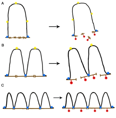
Figure 1. A self-stabilizing tensegrity model for DNA-NM interactions in the cell nucleus as a function of age. (A) In a newborn cell NM
proteins are in a compacted immature state (brown), thus the NM contact
surface is reduced and so a large DNA loop (black) is anchored to two NM
segments by means of two MARs that became actual LARs (blue circles) while
three potential MARs (yellow circles) cannot attach to the NM due to steric
hindrance and lack of enough contact surface. During mitosis biochemical
modification of NM proteins (e.g., phosphorylation, red circles) cause
disassembly of the NM network leading to disappearance of the cell nucleus.
(B) In an adult cell the NM proteins are in a more extended state
offering a larger contact surface, thus further potential MARs become
actualized as LARs reducing the average DNA loop size and increasing the
DNA-NM interactions. Yet phosphorylation of NM proteins leads to nuclear
disassembly during mitosis. (C) In a senescent cell the NM proteins
are fully extended thus offering enough contact surface for several
potential MARs to become actualized as LARs since steric hindrance is
further reduced. DNA loops become shorter on average and DNA-NM
interactions are significantly more numerous. Phosphorylation of NM
proteins during mitosis cannot lead to nuclear disassembly since the
DNA-loops keep separate NM segments bound together and stabilized by means
of the LARs attached to the NM. Thus the available energy becomes limiting
for disassembling the nucleus and the cell cannot enter or perform mitosis.
Throughout the years only a very limited
number of specific proteins have been identified that participate in binding of
DNA to the NM in a sequence-specific fashion [40], such proteins are likely to
be involved in transient-functional DNA-NM interactions. However, given that
there are no MAR consensus sequences and yet the structural DNA-NM interactions
occur on a grand scale (for example, saturation experiments indicate the
existence of some 150,000 salt-resistant DNA binding sites per NM, [56]), these
facts imply that such interactions are the result of indirect readout effects
between DNA and NM proteins thus not equivalent to the direct readout
interactions between transcription factors and specific DNA-functional groups.
Such protein-DNA indirect readouts depend on DNA shape (that is also dependent
on nucleotide sequence) and overall DNA mechanical properties [57]. Thus,
within the eukaryotic genome there are non-coding sequences with a broad range
of affinities for potential attachment sites at the NM, as well as in the NM
there are structural proteins with a broad range of affinities for potential
MARs. A model for explaining how such mutual affinities are regulated and
actualized suggests that the binding of MARs to the NM will depend on three
basic factors: first, the degree of mutual affinity between the DNA sequence
and the potential NM attachment site. Second, the degree of steric hindrance
imposed by the relative density of potential attachment sites per unit length
of NM and the limited deformability (stiffness) of the DNA resulting from its
persistence length [55]. Such a persistence length is actually dependent on
nucleotide composition [58]. Third, the degree of structural stress along the
DNA fibre that modulates the overall deformability of DNA that is a compromise
among bending, opening, uncoiling or breaking up. Hence the very same DNA
sequence can be stably attached to the NM or not depending on the three
above-mentioned factors.
Currently there is ample
evidence that the cell is a high-wired system able to transduce mechanical
information. Indeed, cells within solid tissues are part of a continuum system
of mechano-transduction that couples the extracellular matrix, with the
cytoskeleton and the cell nucleus [59]. Thus the cell can be modelled as a
vector field in which the mechanically linked cytoskeleton-nucleoskeleton may
act as coordinated transducers of mechanical information [55]. The concept of
tensegrity defines structures composed by continuous tension elements and
discontinuous compression elements, in such systems the role of the compression
elements is minimized and the force is distributed among tension elements that
can be slender and lightweight [60]. There is plenty of experimental evidence
that both cell and tissue tensegrity are a biological fact [61,62].
Accordingly, some models predict that permanent changes in cell shape must lead
to modified mechanical interactions within the cell and this would lead to
structural changes within the cell nucleus resulting in redefinition of DNA
loop domains [55]. This has been demonstrated in vitro by inducing a stable
modification in cell shape that resulted in the establishment of new high-salt
resistant DNA-NM interactions and the elimination of some of such previous
DNA-NM interactions, suggesting that both cellular and nuclear shape may act as
cues in the choice of potential MARs that should be actualized as LARs [63].
Evidence for a structural basis for
replicative senescence
The naked DNA loops plus the NM constitute
a "nucleoid" and since the loops remain attached to the NM they are
topologically constrained and supercoiled even after complete extraction of
histones and other chromatin proteins [64,65]. The loop DNA supercoiling is
higher in the regions closer to the NM, save for the actual LARs that
apparently work as buffers against extreme supercoiling [66]. Supercoiling is a
structural barrier against the action of endo-nucleases that hydrolyse the DNA
backbone by a single-strand cleavage mechanism such as DNase I [67]. Also, in
such nucleoids the regions of DNA located close to the NM are relatively
protected from endonuclease action by being immersed in the matrix framework
that may act as a physical obstacle [44,68]. Digestion experiments with DNase
I using nucleoids from freshly isolated rat hepatocytes indicated a progressive
slow-down in the kinetics of nucleoid-DNA digestion as a function of animal
age. This suggests that a larger fraction of nuclear DNA gets closer to the NM
with time [52]. On the other hand, titration with increasing concentrations of
the DNA-intercalating agent ethidium bromide (EB) monitors both the integrity
and supercoiling of the DNA loops [64,69]. The EB acts as a molecular lever
causing the unwinding of loop DNA that produces a halo that surrounds the NM,
this process induces tearing forces as the DNA rotates and expands during
unwinding, such forces impinge upon the NM as DNA is anchored to it. In nucleoids
from newborn (P0) and baby (P7) rat hepatocytes the forces liberated by the
EB-induced DNA unwinding lead in the first case to complete disintegration and
in the second to severe fracturing of the NM framework. However, in nucleoids
from young adult (P80) and senescent (P540) rat hepatocytes the EB releases the
DNA loops creating well-defined DNA halos that surround the undisturbed NM
framework, yet the average halo size is significantly reduced with age. The
average DNA halo size has been correlated with the average DNA-loop size [68,70], and so it was possible to estimate that the hepatocytes from senescent
rats have on average a smaller DNA-loop size than in young adult rats: 31 and
48.9 kbp from tip to base, respectively [52].
The protein composition of the rat
hepatocyte NM shows no significant qualitative difference as a function of
animal age. However, the NM undergoes quantitative changes of the major
constituent proteins such as lamins A, B and C, as well as changes in the
ratios of such proteins [52,71], this is consistent with previous studies
comparing the NM of young and aged human fibroblasts, using 2D electrophoresis [72].
The noticeable increase with age of the three nuclear lamins seems to be
relevant to the obvious strengthening of the NM with age [52]. Indeed,
micromanipulation methods show that nuclei in human embryonic stem cells are
highly deformable and stiffen 6-fold through terminal differentiation, while
nuclei from adult stem cells possess and intermediate stiffness. Knocking down
lamin A/C in differentiated epithelial cells leads to nuclear deformability
similar to that of the adult stem cells [73].
There is an average increase
in diameter and volume of both the nucleus and the NM in hepatocytes from
senescent rats (P540) and so the NM framework becomes proportionally larger
with age [52], this correlates with the reported increase in nuclear roundness
with age that smoothes out the invaginations of the nuclear contour [72]. The
mean compactness of the NM proteins decreases during development being in the
adult rat one fourth of that in the 16-day foetus, suggesting that the NM
protein network becomes more extended as development progresses [71]. Such a
reduction of NM-protein compactness suggest the progressive shift from a
nuclear substructure consisting of unconnected, merely clustered
ribonucleoprotein (RNP) fibres and granules to a mature, continuous internal
network consisting of interconnected and branched RNP filaments that connect to
the nuclear lamina [39,74,75]. A reduction in NM protein compactness or
smoothing-out of NM curvature, together with an increased NM volume would also
make available a larger contact surface for potential MARs that may bind to the
NM depending on their affinity for NM proteins and the degree of local steric
hindrance resulting from compactness or extension of the NM protein framework.
The HLMCA model proposes that eudomains are the default state for chromatin and
that the epigenetic heterodomains are metastable and thus prone to decay into
less folded chromatin structures [37] and so there is evidence that
heterochromatin is much reduced in the nuclei of aged cells [35,36]. Thus, in
nuclei from aged cells virtual MARs formerly occult within heterodomains may
become available for interacting with the NM establishing further DNA loops.
The following observations: reduction of
loop DNA sensitivity to DNase I, reduction of the average DNA-loop size,
increase in the nuclear volume and reduction of nuclear deformability with age [52,73], correlate with the known reduction of the cell proliferating potential
with age, even when cells have not undergone repeated cell division cycles
through the years (as in the case of quiescent rat hepatocytes, [33]). Thus, the
experiments with rat hepatocyte nucleoids indicate that in nuclei from aged
animals there is a larger number of DNA-NM anchoring interactions, resulting in
a larger number of DNA loops that are significantly shorter and more stable
than those in nuclei from younger-animal cells, and so the actualization of
potential DNA-NM interactions increases with time.
An important question is what could be the driving
factor behind the post-natal increase in nuclear size and volume that
establishes the basic condition for further consolidation of the DNA-NM
interactions. A typical feature of senescent cells is that they are large-sized
(hypertrophic), also it is well known that cell size increases in culture as
cells progress toward senescence [76]. Moreover, the liver is an organ that
keeps growing during the post-natal period but all evidence suggests that this
growth is primarily by hepatic hypertrophy that correlates with a trend of the
hepatocytes to undergo polyploidization as a feature of cell maturation. In the
liver of normal young rats already 60% of hepatocytes are mononucleated
polyploid cells [77], indicating that DNA synthesis has been proceeding in
absence of both karyokinesis and cytokinesis; and in older rats there is a
direct correlation between higher prevalence of polyploid cells and increasing
age [78]. There is evidence that the onset of polyploidy in hepatocytes is
associated with weaning and assumption of independent feeding in rodents and
that the insulin/Akt pathway is involved in the control of this process [78,79].
Interestingly, the nutrient-sensing TOR pathway that is activated by insulin,
growth factors and nutrients is an essential controller of cell growth. TOR
(target of rapamycin) is a serine/threonine kinase that participates in two
distinct multiprotein complexes (TORC1 and TORC2) each of which signals through
a different set of effector pathways. TOR is conserved from yeast to human and
strikingly the inhibition of the TOR pathway prolongs lifespan in yeast, worms,
flies and mice [80,81]. Thus it was predicted that blocking the cell cycle
without a corresponding block of cell growth would cause cell senescence and
this has been experimentally confirmed in vitro since when the TOR pathway was
active and the cell cycle was blocked cellular senescence occurred [82]. This
important result ties in with recent evidence in vivo that the insulin/Akt
pathway directly or indirectly through TORC2, is involved in the process that
leads to generation of polyploid hepatocytes in rodents [79], suggesting that
growth and aging may share a common molecular mechanism [83].
From the structural
perspective, the topological organization of higher-order DNA structure based
on selective use of a limited set of potential MARs (as seen in nuclei from
newborn and baby animals) is highly asymmetrical and the natural trend for most
physical systems is towards reducing the asymmetries in such a way that the
system evolves in time so as to become more symmetrical [84-86]. A topological
configuration in which most potential MARs are actually bound to the NM, thus
resulting in shorter and more stable DNA loops, is also a more symmetrical
structural attractor. Moreover, since entropy is not a measure of disorder or
chaos, but of energy diffusion, dissipation or dispersion in a final state
compared to an initial state [87], such a highly-stable DNA-loop configuration
satisfies the second law of thermodynamics since the structural stress along
the DNA molecule is more evenly dispersed within the nuclear volume by
increasing the number of DNA-NM interactions (thus increasing, in terms of
molecular thermodynamics, the occupancy of more microstates in phase space). A
larger number of DNA-NM interactions create a structural complex, similar to a
hanging bridge in which beams (proteins) and tensors (DNA) interact for
creating a highly stable overall structure. Thus any relatively stable
interaction between two NM-protein filaments will be further stabilized if a
given DNA loop interacts with both filaments, but also the stability of the DNA
loop shall be increased by the interaction with both protein filaments,
resulting in a self-reinforcing structural stability that operates at the scale
of the whole interphase nucleus (Figure 1).
However, there are some terminally
differentiated cells whose post-mitotic stage is rather short-lived (in the
order of days). Indeed, for such cells terminal differentiation is the
antechamber of cell death. Such is the case of lymphocytes, neutrophils, sperm
cells or epidermal cells, all of which have very limited life spans after terminal
differentiation and either do not constitute solid tissues or are located close
or at the open edge of a solid tissue. In such cells there is limited scope for
tissue mechano-transduction acting as guide for nuclear organization.
Interestingly, in these cells terminal differentiation is linked to induction
of DNA strands breaks that preferentially occur at sites involving MARs,
liberating DNA fragments of some 50 kbp, that roughly correspond to the average
distribution of chromatin looped domains [88]. Indeed, ribo-nucleoprotein-masked
nicks exist in the genome distributed on average every 50 kbp, suggesting that eukaryotic genomic DNA is composed of contiguous
rather than continuous single strands, interrupted at the boundaries of
interphase chromatin loops [89]. This fact supports the notion that attachment
to the NM contributes to stabilize the long-range DNA structure. On the other
hand, massive breaking of DNA in regions corresponding to actual LARs would
cause inability to perform appropriate chromosome condensation during mitosis
as well as to complete nuclear reassembly. Several important processes of
nuclear physiology, such as replication, transcription and processing of
primary transcripts occur at macromolecular complexes located upon the NM [90-92].
Thus the topological relationship between DNA loops and the NM is very
important for appropriate nuclear physiology. For example, productive infection
by herpes simplex virus type 1 induces DNA breaks and wholesale alteration of
higher-order structure of the host cell chromatin, resulting in loss of
DNA-loop supercoiling and organization that correlates with complete inhibition
of host-cell replication and transcription [69,93-95]. Indeed, correct repair
of DNA damage must include the recovery of both the double helix integrity and
the complex third-dimensional DNA topology, otherwise the cell will not survive
[96,97]. Therefore, cells with overall disruption of higher-order DNA
structure are irreversibly committed to functional failure in the short term.
Why a stable higher-order nuclear
organization leads stochastically to replicative senescence
Highly stable
physical systems are quite resistant to change and have a much reduced dynamic
potential. Thus, a structurally-stable cell nucleus would not be the seat of
both the dynamic transitions necessary for mitosis and the rearrangements of
chromosome territories and chromatin domains in early G1 [98] that normally
occur in cells with a positive proliferating potential, since the energy cost of
nuclear disassembly and reassembly will be limiting for the cell. Indeed, the
sub-nuclear organization of interphase chromosomes in pre-senescent mammalian
cells is quite different from that in proliferating or quiescent cells,
indicating that on average the spatial organization of the genome within the
nucleus changes with age [99]. Thus the nuclear higher-order structure
established by the topological interactions between chromatin and NM
constitutes an integral structural system that naturally but relentlessly
evolves towards a more symmetrical and highly stable state. Since this process
obeys thermodynamic constraints it must follow a stochastic behaviour that
nevertheless increases its probability as a function of time. This might be a
more general, physical basis for terminal, non-reversible cell differentiation,
leading to cellular replicative senescence and a long-lasting, highly stable
post-mitotic state that is independent of the action of soluble factors acting
in trans and that occurs in a stochastic but time-dependent fashion
within cell populations, whether or not the affected cell has previously
divided (thus independently of any cell-division counting mechanism).
Heterochrony is
developmental change in the timing of events, leading to changes in size and
shape. There is no doubt that during embryogenesis there are changes in the
rate or timing of development of some cell lineages in the body relative to
others, so that different cell lineages develop at different rates.
Mechano-transduction during tissue morphogenesis may induce changes in the
differentiation state of cells and such a modification of the differentiation
state also impinges on the potential morphogenetic trajectory by limiting the
repertory of changes in cellular size and shape. Heterochrony may alter the
distribution of probabilities of stochastic events such as the rate of
actualization of DNA-NM interactions, hence some cell types such as neurons
reach terminal differentiation and became post-mitotic earlier than others, depending
on their morphogenetic trajectory. As a corollary it can be concluded that such a highly-stable nuclear
post-mitotic structure cannot be altered, reverted or bypassed by any known
oncogenic stimuli and as such is the true barrier against tumorigenesis.
This work was
sponsored by CONACYT grant 48447-Q and UAEMéx, grant 2212/2006. I thank Dr.
Alejandro Martínez-Gómez for drawing the figure.
The authors in this
manuscript have no conflict of interests to declare.