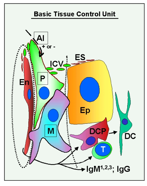
Figure 1.Schematic drawing of the basic "tissue control unit," which consists of monocyte-derived cells (marked M in the figure), vascular pericytes (P), and autonomic innervation (AI, dashed arrow), and the involvement of other components of the tissue control system (solid arrows). Monocyte-derived
cells physically interact with adjacent epithelial (Ep) and
endothelial cells (En) through the basement membranes (dotted lines),
and influence pericytes, which secrete intercellular vesicles
(ICV). These vesicles collapse into the so-called empty spikes (ES)
releasing their content (growth factor/cytokine) after reaching target
cells. The activity of pericytes is stimulated or inhibited by
autonomic innervation (+ or -) which controls quantitative aspects
of tissues. Interaction of MDC with endothelial cells may stimulate
homing of T lymphocytes (T) and monocyte-derived dendritic cell precursors
(DCP; also known as veiled cells) differentiating into mature dendritic
cells (DC). The dendritic cell precursors and T cells interact themselves
and stimulate advanced differrentiation of epithelial cells.
IgMs regulate early (IgM1), mid (IgM2), and late differentiation
(apoptosis) of epithelial cells (IgM3), and IgG associates with aged cells
(see Figure 2 and 3). The monocyte-derived cell system (including
intraepithelial DCP and mature DC) is postulated to play a dominant role
in the regulation of qualitative aspects of tissue-specific cells, including
expression of ligands for intraepithelial T cells and regulating autoantibody action.
Monocyte-derived cells also carry "stop effect" information (Figure 10B),
presumptively encoded at the termination of immune adaptation (Figure 10A),
which determines the highest state of epithelial cell differentiation allowed
for a particular tissue. For details see Ref. [3,4,33]. Reprinted from Ref. [4], © Antonin Bukovsky.