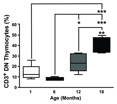
Figure 1.CD3 expression on DN thymocytes shows an age-dependent increase. Thymocytes from different aged
mice were stained with anti-CD3, anti-CD4 and anti-CD8 mAb, analysed by
flow cytometry and CD3 on DN cells was determined gating the appropriate
population. This study revealed that the proportion of CD3+ DN
thymocytes showed an age-dependent increase. (One month n=5; six months
n=5; 12 months n=8; 18 months n=4). *P<0.05; **P<0.01;
***P<0.001.