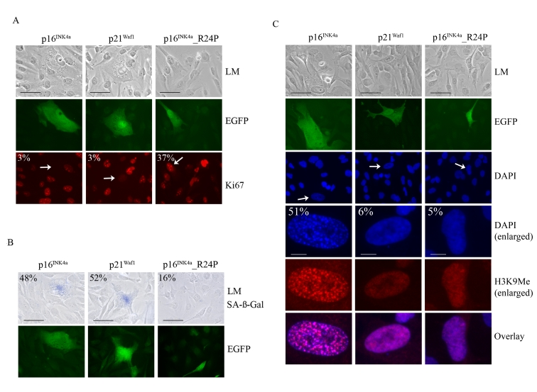
Figure 4.Impact of p16 INK4a or p21Waf1 expression on the cellular senescence program. WMM1175 melanoma
cells were cotransfected with plasmids encoding p16INK4a, p21Waf1
or the melanoma-associated p16INK4a_R24P along with pCMV-EGFPN1,
which was used as a marker of transfection. Five days post
transfection cells were fixed, permeabilized and analyzed. (A) Cell
proliferation was monitored by Ki67 immunostaining and the percentage of
transfected WMM1175 cells with positive Ki67 staining is indicated and was
determined from at least two separate transfection experiments and from a
total of at least 300 cells. All standard deviations were less than ±5% (bar=100μm). (B) Transfected WMM1175 cells
were analyzed for SA-β-Gal
activity, and the percentage of positive SA-β-Gal transfected cells is
indicated, and was determined as detailed above (bar=100μm). (C) The appearance of
SAHF was analyzed by immunostaining with antibodies to H3K9Me and
co-staining DNA with DAPI. The percentage of transfected cells with detectable
foci is indicated, and was determined as detailed above (bar=100μm).