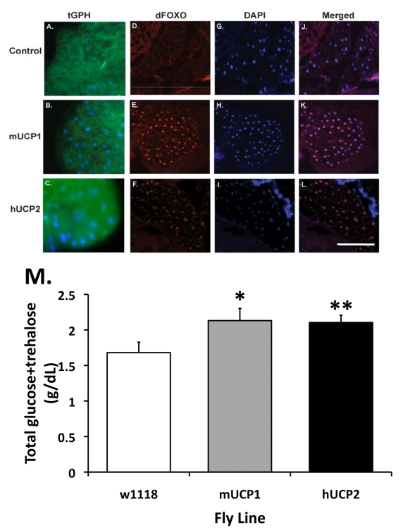
Figure 2.Normal and reduced systemic insulin signaling as reflected by the cellular localization of the PH-tagged GFP reporter protein (tGPH) and dFoxO in fat body cells.(A-C)
Increased UCP activities in the adult IPCs attenuate systemic insulin
signaling events. In control flies, under normal growth conditions and a
full strength PI-3' kinase activity, tGPH is predominantly located at the
plasma membrane of each fat body cell (A). (B-C) UCP
expression in adult IPCs results in a diffused, cytoplasmic distribution of
the tGPH protein. Control: dilp2-Gal4, tGPH; mUCP1: dilp2-Gal4/UAS-mucp1, tGPH; hUCP2: dilp2-Gal4/UAS-hucp2, tGPH. (D-F)
Increased accumulation of dFoxO in the nucleus of pericerebral fat body
cells in adult dilp2-Gal4/UAS-mucp1 and dilp2-Gal4/UAS-hucp2 flies indicates
reduced insulin signaling. Cryosections of adult heads were stained with
an α-dFoxO antibody
followed by Alexa 568-conjugated secondary antibodies. A strong nuclear
staining of the dFoxO protein was observed in the pericerebral fat body in
both dilp2-Gal4/UAS-mucp1 (mUCP1, Panel
E) and dilp2-Gal4/UAS-hucp2 (hUCP2, Panel
F) flies but not in dilp2-Gal4/w1118 (Control, Panel
D) flies. All sections were counter stained with DAPI (Panels G-I) to
locate the nucleus of each cell. Merged images of anti-FoxO staining and
DAPI are shown in Panels J-L. (M) Elevated levels of fasting
circulating sugars are measured in adult dilp2-Gal4/UAS-mucp1 and dilp2-Gal4/UAS-hucp2 flies. An
average of 29% increase in circulating sugars measured in 14-day-old dilp2-Gal4/UAS-mucp1 (mUCP1) and dilp2-Gal4/UAS-hucp2 (hUCP2) females
as compared to control dilp2-Gal4/w1118 (w1118)
females. Each bar represents mean +
SEM. N=5-7, *p= 0.046, **p=
0.05 (Student's t test).