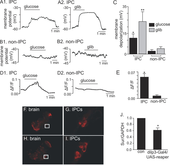
Figure 4.Electrophysiological, Ca 2+ influx, and expression evidence of functional KATP channels in adult IPCs.(A1-C)
Membrane depolarization of adult IPCs in response to glucose and
glibenclamide. (A1) Trace of membrane potential from an adult IPC
in the whole brain preparation shows that exposure to high glucose (80 mM)
evoked a reversible membrane depolarization. (A2) Trace of membrane
potential from an adult IPC shows that exposure to a commonly known KATP
channel blocker, glibenclamide (glib, 20 μM) also evoked a reversible
membrane depolarization. (B1 and B2) traces of membrane
potential from adult non-IPCs show that these cells do not respond to
glucose or glibenclamide. (C) Average membrane potential response
to glucose and glibenclamide of IPCs (N=5) and non-IPCs (N=3). Glucose (*)
and glibenclamide (**) significantly increased membrane potential of IPCs
as compared to non-IPCs, *p and **p <0.05 (Student's t test). Each
bar represents mean +
S.E.M. (D1-E) Glucose-dependent Ca2+
influx measured in adult IPCs. (D1) Normalized fluorescence trace (∆F/F) (see
Materials and Methods) recorded from an acutely dissociated adult IPC shows
that exposure to glucose (80 mM) increased fluorescence intensity, thus
indicating an increase in intracellular Ca2+. (D2)
Normalized fluorescence trace (∆F/F) recorded from an acutely
dissociated non-IPC shows that glucose does not increase intracellular Ca2+
in these cells. (E). An average of normalized fluorescence
intensity in response to glucose demonstrates a significant increase in Ca2+
influx recorded from IPCs (N=6) as compared to non-IPCs (N=3). *P= 0.007
(Student's t test). Each
bar represents mean +
S.E.M. (FI) In situ hybridization
of whole mount adult brains demonstrates dSur expression in
IPCs (F-G) when probed with anti-sense dSur probes and dilp2 expression (HI)
when probed with anti-sense dilp2 probes. The
IPCs marked in squares are shown in panel G for dSur signals and
panel I for dilp2
signals.
(J) Quantitative real-time PCR analysis reveals an average of 33%
reduction in dSur transcripts
when the IPCs are partially ablated using an IPC-specific driver, dilp3-Gal4 to drive the
expression of a pro-apoptotic gene, reaper. The
housekeeping gene GAPDH was used as the reference gene. Each bar
represents mean +
S.E.M. N=5, *p<0.001 (Student's t test).
Control: dilp3-Gal4/w1118.