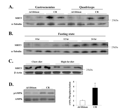
Figure 3.Diet-sensitive expression of SIRT3 and AMPK in muscle tissue.(A) Mice
were fed NIH-31 standard feed ad libitum or NIH-31/NIA-fortified
diet (Harlan Teklad) with a daily food allotment of 60% of the control mice
to establish caloric restriction (CR); twelve months after the onset of CR,
tissues were harvested to examine SIRT3 expression. (B) Mice were
deprived of food for 24 hours, and SIRT3 level in EDL muscle was determined
by Western blot analysis. (C) SIRT3 protein expression is decreased
in murine hind-leg muscle after 3 months of high-fat diet feeding; total
hind-leg tissue protein was isolated and analyzed. (D) AMPK T-172
phosphorylation and AMPK total protein in the quadriceps of the caloric
restricted mice were assayed; AMPK phosphorylation was determined as
phospho-AMPK normalized by total AMPK. N=3, *P < 0.05.