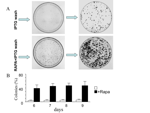
Figure 6.Clonal proliferation of competent cells.HT-p16 cells
were plated in 100-mm plates. The next day, 50 μM IPTG with or without
rapamycin, if indicated (RAPA), was added. After 3 days, the plates were
washed to remove IPTG and RAPA. (A) Photographs. Upper panel: On
days 5 and 8 (after IPTG removal), plates were fixed, stained and
photographed. Lower panel: On days 5 and 8 (after IPTG removal), plates
were fixed, stained and photographed. (B) Number of colonies. On
days 6, 7, 8 and 9 (after IPTG removal), plates were fixed, stained and
photographed. The number of colonies was counted and results are shown as
percent of plated cells in log-scale.