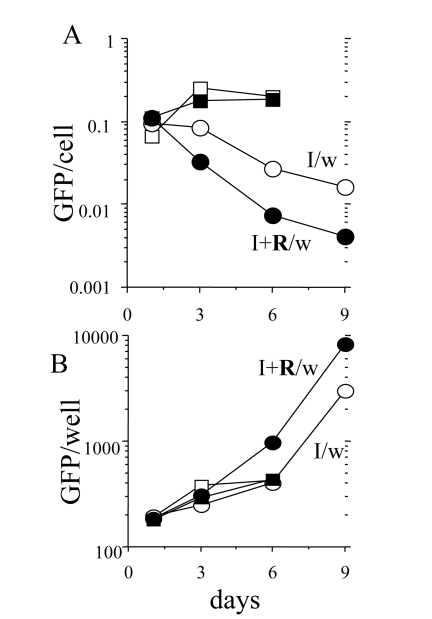
Figure 8.Loss of hypertrophy during proliferation of competent cells.500 HT-p21 cells
were plated in 12 well plates. The next day, either IPTG alone or IPTG plus
rapamycin were added. After 3 days, plates were washed (I/w and I+R/w) or
left unwashed. GFP per well was measured and cells were counted at days 1,
3, 6 and 9. GFP per cell was calculated (upper panel). Results are shown in
arbitrary units (M±m). Open and closed squares: IPTG and IPTG plus Rapa,
respectively. Open and closed circles: IPTG washed (I/w) and IPTG plus Rapa
washed (I+R/w), respectively. When cells resumed exponential proliferation,
GFP per cell dropped to normal levels. Due to robust proliferation, there
was an increase of GFP per well.