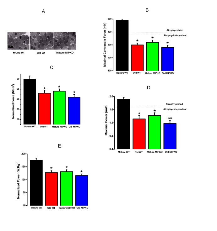
Figure 2.Evidence for muscle atrophy, decreased contractile force, and reduced power in skeletal muscles suggested similarity from old WT and MIPKO mice. In all figures,
the black bars are mature, wild type mice, the red bars are old, wild type
mice, the green bars are mature MIPKO mice, and the blue bars are old,
MIPKO mice. (A) Typical Toluidine blue-stained cross sections of
EDL muscles from young Wt, old Wt, and mature MIPKO mice. The cross-sectional
areas of old Wt and MIPKO cells are significantly reduced compared with those of
the young Wt. (B) Maximal contractile force in EDL muscle for each genotype.
Atrophy (decrease in muscle cross-sectional area) can explain ~ 1/2 of the drop
in total force (note the dotted horizontal line), but does not account for the
complete decrease in contractile force. (C) Data from B, except
that force is normalized per cross-sectional area (N/cm2). This figure illustrates
the atrophy-independent component of contractile dysfunction. (D) Maximal
power in EDL muscle from all four animal models. (E) Data from panel D
was normalized per cross sectional area of muscles. It shows that a significant drop
in power is atrophy-independent. Data is the average ± SE of 24 EDL muscles from 12
mice for each genotype. * indicates a significant difference (p < 0.01) between the
control muscles and a particular genotype. ** indicates a significant difference
(p < 0.01) between the old MIPKO mice and the old Wt and mature MIPKO mice.