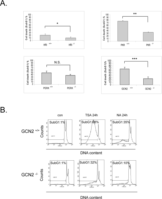
Figure 4.eIF2α kinases enhance sensitivity to vorinostat independently of eIF2α phosphorylation.(A) The indicated MEFs were treated with DMSO (con) or 10 μM vorinostat (SAHA) for the different time periods (HRI MEFs 72h, PKR and PERK MEFs 48h, GCN2 MEFs 24h). Cells were subjected to FACS analysis after propidium iodide staining. Cell death is represented by the percentage (%) of cells in SubG1. Histograms represent the mean cell death from three independent experiments for the corresponding time periods (N=3, treated minus untreated). Bars denote S.E.M.. Statistical significance of the differences as calculated by Student's t-test is with *P<0.03 **P<0.004, ***P<0.005. N.S. = not significant. (B) GCN2 +/+ and GCN2 -/- MEFs treated with DMSO (con), 1 μM trichostatin A (TSA) or 50 mM nicotinamide (NA) for 24h. Cells were subjected to FACS analysis after propidium iodide staining. Cell death is represented by the percentage (%) of cells in SubG1.