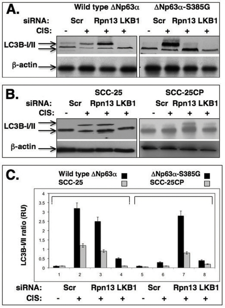
Figure 5.Cisplatin induces the autophagic process through a LKB1 up-regulation(A) Wild type ΔNp63α cells and ΔNp63α -S385G cells and (B) Sensitive (SCC-25) and resistant (SCC-25CP) squamous carcinoma cells were exposed to control media and 10μg/ml cisplatin for 24h. Cells were transiently transfected with scrambled siRNA, siRNA against Rpn13 or LKB1. Cells were grown up in the presence of lyzosomal protease inhibitors (10 μg/ml of both E64d and pepstatin A). Protein levels of autophagic markers were analyzed by immunoblotting with indicated antibodies. β-actin expression was used as a loading control. (C). Quantitative analysis of LC3B -I/II ratio. Immunoblots were scanned using PhosphorImager (Molecular Dynamics) and quantified by Image-Quant software version 3.3 (Molecular Dynamics). Values of LC3B-II were expressed as a portion of LC3B-I values defined as 1. The LC3B-II/LC3B-I ratios were plotted as bars using the Microsoft Excel software with standard deviations (± SD, p>0.05) resulting from three independent experiments and three individual measurements of each experiment. Black bars represent the set of wild type ΔNp63α/ΔNp63α-S385G cells, while grey bars represent a set of SCC-25/SCC-25CP cells.