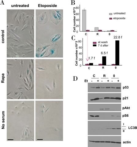
Figure 4.Effects of rapamycin and serum starvation on etoposide-induced senescence in RPE cells.A-C. RPE cells were plated at 5000/well in 12 well plates, and the next day either treated with 10 nM rapamycin in complete medium (R), or placed in serum-free medium (no serum or 0), or left in complete medium (control). The next day, 0.5 μg/ml etoposide (Et) was added, as indicated.
B. After 4 days, cells were stained for beta-Gal and micro-photographed (bar - 50 micron)
C. Proliferative potential. In replicate plates, cells were washed and incubated in complete, drug-free medium for 6 days and then counted (black bars). Note: red bars corre-spond to red bars in panel B. Fold (f) increase in a cell number after drug removal.
D. Immunoblot. Cells were plated in 6 well plates. The next day, cells were treated with 0.5 μg/ml etoposide (Et) for 24 hrs: control -C, rapamycin -R, no serum −0.