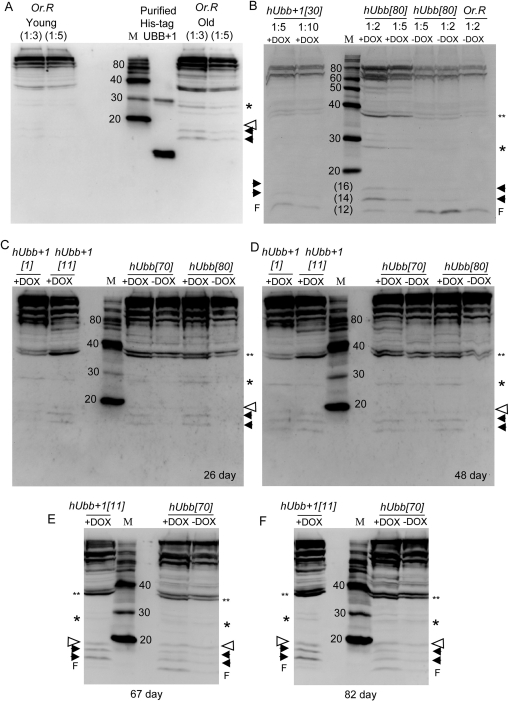
Figure 4.Western blot analysis using antibody specific for hUbb+1Total protein was isolated from 30 male flies of the indicated genotypes, and 1/8 of the sample was assayed for the presence of protein that would be recognized by hUbb+1 antibody. Where indicated protein samples were diluted 1:2, 1:3, 1:5 or 1:10 to confirm sensitivity of the assay to relative protein concentrations. In panels B-F all samples are diluted 1:3. (A) Molecular weight markers were run alongside His-tagged hUbb+1 purified from E. coli cells as well as total protein isolated from 30 “young” (10 day old) and “old” (65 day old) male Oregon-R control flies, as indicated. (B) “Young” (10 day old) flies of the indicated genotypes. Note the hUbb[80] –DOX sample lanes contain cross-reacting material that is unresolved from the gel front (F), and is interpreted as degradation products. This material was not present in other hUbb[80] protein samples (see panels C and D). (C) Flies cultured +/− DOX for 26 days. (D) Flies cultured +/− DOX for 48 days. (E) Flies cultured +/− DOX for 67 days. (F) Flies cultured +/− DOX for 82 days. Where visible the gel protein front (F) is indicated. Solid arrowheads indicate two species of <20Kd, either of which might represent Ub+1 monomer, which has an expected size of ~11Kd. Open arrowhead indicates species at expected position for Ub+1 ligated to one Ub wild-type protein (~11Kd + ~8.5Kd = ~19.5Kd). Single asterisk indicates species at expected position for Ub+1 ligated to two Ub proteins (~ 11Kd + ~17Kd = ~28Kd). Double asterisk indicates species at expected position for Ub+1 ligated to three Ub proteins (~11Kd + ~25.5Kd = ~37Kd). Estimations of sizes of various species are presented in Supplemental Materials.