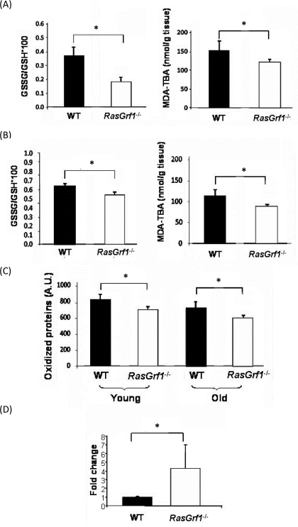
Figure 3.Decreased Oxidative Stress in RasGrf1−/− Mice(A) Oxidative stress was measured in brain from 4-6 months old animals. Glutathione redox ratio was significantly higher in WT (n=7) than in RasGrf1−/− male mice (n=8). Malondialdehyde (MDA) levels were significantly lower in brain of RasGrf1−/− mice (n=8) than in that of WT (n=7). MDA was measured by the formation of the aduct malondialdehyde-thiobarbituric acid (MDA-TBA) (*p<0.05). (B) Oxidised glutathione and malondialdehyde levels were significantly lower in liver of RasGrf1−/− male mice. n for WT = 7 and for RasGrf1−/− = 8 (*p<0.05) (4-6 months old). (C) Oxidised proteins in young (4-6 months old) and old (20-22 months old) WT mice were significantly higher than in RasGrf1−/− male mice (*p<0.05). These were measured by western blot with antibodies directed against aldehydes in proteins. (D) Cytochrome c oxidase expression is significantly higher in RasGrf1−/− than in WT male mice of 4-6 months old (*p=0.037). Total RNA from selected tissues was isolated and used to quantify cytochrome c oxidase by real time RT-PCR.