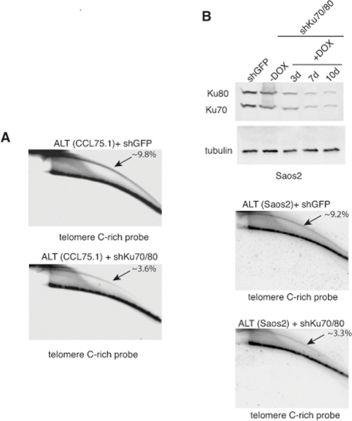
Figure 4.Depletion of Ku70/80 reduces the levels of t-circles in CCL75.1 and Saos2 cells(A) Genomic DNA isolated from CCL75.1 cells expressing shRNAs targeting Ku70/80 or GFP for 7 days was digested with HinfI and RsaI, separated by 2DGE, blotted, and probed with a telomeric (CCCTAA)4probe. The samples shown in each panel were run and processed in parallel under the same hybridization and washing conditions. The approximate levels of t-circles present in each sample (expressed as a percentage of the total telomeric DNA) were estimated as in [14] and is shown in the upper right corner. (B) (top panel) Saos2 cells were infected with lentiviruses for the conditional expression of shRNAs for Ku70and Ku80 or GFP and analyzed either before or 3, 7 or 10 days after the addition of 1.0 mg/ml DOX to the media. The levels of Ku70 and Ku80 were determined by Western blotting with antibodies against Ku70 and Ku80. We estimated that Ku70/80 shRNAs reduce Ku70/80 expression ~80% compared to GFP shRNAs control. Antibody against tubulin was used as a loading control. (middle and bottom panels) DNA isolated from Saos2 cells 7 days after DOX addition was digested with HinfI and RsaI, separated by 2DGE, blotted, and probed with a telomeric (CCCTAA)4probe. The samples shown in each panel were run and processed in parallel under the same hybridization and washing conditions and the approximate levels of t-circles present in each sample (expressed as a percentage of the total telomeric DNA) is shown in the upper right corner.