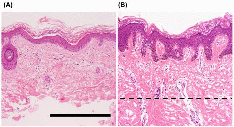
Figure 1.Representative histological features of the skin from patients with Werner syndrome (WS)Representative images of skin specimens from WS patients, stained with hematoxylin and eosin. The left panel (A) shows a sample of skin after removal of dermal tissue with a scalpel, and the right panel (B) shows a sample before such treatment. Dashed line indicates the treatment border. Figures A and B show microscopic views of skin from the ankle of a 43-year-old man (WS-2) and the lower leg of a 41-year-old man (WS-7), respectively. Remarkable atrophy of the epidermis and dermis is evident. Mild hyperkeratosis, mild dermal hyalinization, and atrophy of the skin appendages (hair follicles) are present in both cases, with marked flattening of the rete ridges in (A). No marked inflammatory cell infiltration is evident in either case. Scale bar, 50 μm.