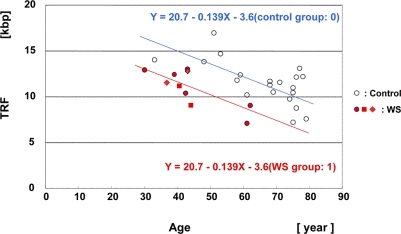
Figure 3.Scatter plot analysis of HinfI-digested TRF length in skin samples from WS patients and controlsMultiple regression analysis yielded a regression line for the 8 WS patients (10 samples) (Y = 20.7 − 0.139X (age) − 3.6 (WS group: 1); in red), and a regression line for the non-WS control subjects, aged between 30 and 80 years (n = 21) (Y = 20.7 − 0.139X (age) − 3.6 (control group: 0); control line denoted in blue). The difference of TRF values between the groups was significant. P = 0.00026. The same patient at the ages of 37 and 43 years (WS-4 case: ◆). The same patient at the ages of 41 and 44 years (WS-7 case: ■).