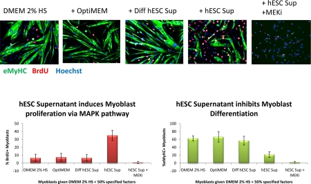
Figure 1B.Primary myoblasts were cultured for 24 hours in DMEM + 2% Horse Serum and 50% of the supernatant specified. 10 μM of MEK inhibitor was added to some wells, as indicated. At 24 hours, cells were pulsed with 10 μM BrdU for 2 hours and fixed with 70% ethanol. Cells were immuno-stained for eMyHC (green) and BrdU (red); Hoechst (blue) was used to label all nuclei Automated imaging of these cells was done using ImageXpress and automated counting of percent of eMyHC+ and BrdU+ cells was performed by quantifying at least 100 sites per experimental sample by MetaExpress. hESC supernatant enhanced myoblast proliferation in a MAPK-dependent manner and diminished differentiation into myotubes.