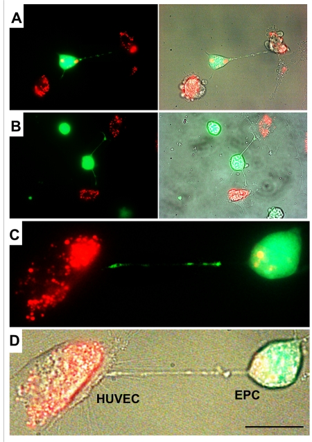
Figure 2.A gallery of images of TNT formation between HUVEC and EPC.(A, B, C and D) Images depict scarce transfer of lysotracker red-labeled lysosomes from stressed HUVEC to EPC labeled with CFDA SE green. Note that multiple TNT exist between two cell types, only a few of them convey lysosomes. Panels C and D depict enlarged fluorescence and bright-field images of HUVEC and EPC. Bars 20μm.