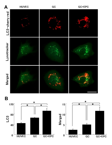
Figure 8.Lysosomal and autophagosomal punctae in control or stressed (GC) HUVEC.(A) Representative images of HUVEC expressing LC3-cherry red (autophagosomal marker, upper raw) and lysotracker (middle raw) under basal conditions, following application of GC in the absence or presence of intact EPC. The lower panel depicts corresponding merged images. Co-localization of autophagosomal and lysosomal markers indicates their fusion (autolysosomal formation), which has been shown to be subverted in stressed HUVEC (Bars 20μm). (B) Quantitative summary of autophagosomal punctae and their fusion with lysosomes (co-localized). Image J software (National Institutes of Health) was used for analysis. Asterisks p< 0.05; n=5.