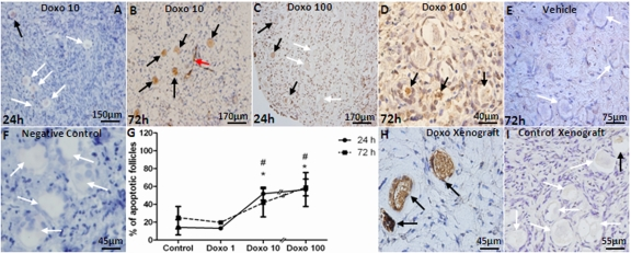
Figure 2.Induction of apoptotic primordial follicle death by doxorubicinAC3-positive (black arrows) and negative (white arrows) primordial follicles and apoptotic vasculature (red arrow) in human ovarian cortical pieces. A-D) Doxorubicin induces apoptosis of human primordial follicles after 24h culture in 10 and 100 μg/ml concentrations. E) Vehicle control at 72h. F) Negative control are shown where primary antibody omitted. G) A significant correlation was observed between the dose of doxorubicin and percentage of apoptotic primordial follicles in cultured OCTs (Spearman's rank correlation coefficient: 0.785; p=0.0001). H) A high number of apoptotic primordial follicles was seen in human ovarian tissue xenografted into the dorsal muscle of SCID mouse after treatment with 10mg/kg doxorubicin. I) Xenogafted vehicle-treated control.
* Significantly different from controls at 24 or 72h.
# Significantly different from 1 μg/ml doxorubicin dose at 24 and 72h.