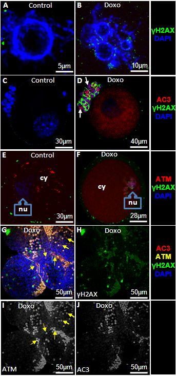
Figure 5.Doxorubicin induces double strand DNA breaks, activates ATM, and causes apoptosis in mouse oocytesTo study the in vivo effect of doxorubicin on mouse ovarian follicles and their cumulus cells, animals were injected with a single dose of doxorubicin 10 mg/kg. GV oocytes and small preantral follicles were retrieved 24h later from ovaries and evaluated by confocal microscopy. A&B) Doxorubicin treatment leads to multinucleation as well as increased expression of γH2AX. C&D) Doxorubicin-induces apoptosis of the oocyte and surrounding granulosa cells (arrows) in association with the induction of DSBs. E&F) Expression and localization of ATM in oocytes after doxorubicin treatment. While a higher amount of ATM is localized in the cytoplasm (cy) of the control oocytes (E), doxorubicin-treated oocytes show higher localization in the nucleus(nu)(F). G) Consistent with its impact on oocytes, ATM expression also increases in granulosa cells of small antral follicles after doxorubicin treatment (yellow arrows). H) Note that doxorubicin treatment induces DSBs in granulosa cells of small antral follicles (Split channel from image G). I) A minority of granulosa cells are non-apoptotic and express ATM indicating that DNA repair might have enabled them to overcome genotoxic stress (yellow arrows) (split channel from image G). J) The majority experience apoptosis despite the activation of ATM (split channel from image G).