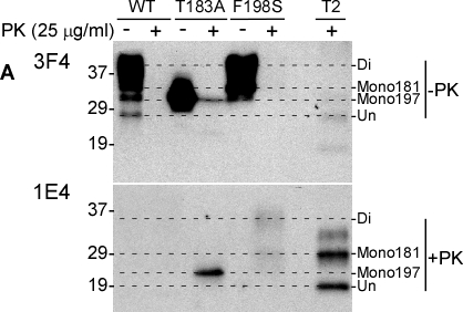
Figure 1.Detection of untreated and PK-treated PrP from three types of cultured cells with 3F4 and 1E4A: Western blotting of cell lysates with or without PK-treatment at 25 μg/ml) probed with 3F4 (upper panel) and 1E4 (lower panel). WT: Lysates of cells expressing PrPWt. T183A: Lysates of cells expressing PrPT183A mutation. F198S: Lysates of cells expressing PrPF198S. T2: PrPSc type 2 control from sCJD. Di: Diglycosylated PrP. Mono181: PrP monoglycosylated at the first site. Mono197: PrP monoglycosylated at the second site. Un: Unglycosylated PrP. Comparison of affinities of 1E4 and 3F4 antibodies to the full-length and N-terminally truncated human PrP. The dashed-lines are used to align PrP bands on the blots.
Immunofluorescence detection of untreated and treated PrP with 3F4 and 1E4. Panels I-III: Cells expressing human PrPWt. Panels IV-VI: Cells expressing human PrPT183A. Panels VII-IX: Cells expressing human PrPF198S. Panels I, IV, and VII: Staining with 3F4. Panels II, V, and VIII: Staining with 1E4. Panels III, VI, and IX: Staining without anti-PrP antibodies. Panels X and XI: Wild-type cells staining with 1E4 before and after PK-treatment. Panels XII and XIII: Wild-type cells staining with 3F4 before and after PK-treatment.