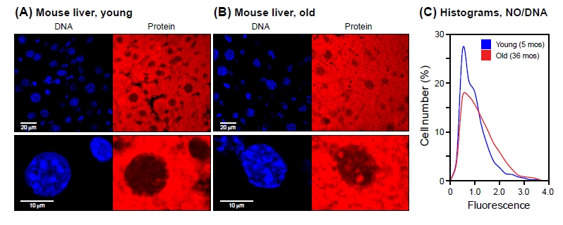
Figure 5.NanoOrange staining of mouse liver tissueCryosections of liver tissue were processed and stained with NanoOrange (NO) as indicated in Methods. (A) Young (5 months old). (B) Old (36 months old). Left panels, DAPI staining; right panels, NanoOrange staining. Lower panels shown selected nuclei at high magnification (note scale bars at the left of the DAPI images). Note bright NO-staining foci in old nuclei that colocalize with DAPI-staining foci. All mice were C57Bl/6 males and were healthy at the time of sacrifice. (C) Histograms of NanoOrange staining for young and old liver tissue. Fluorescent images were quantified using CellProfiler software as indicated in Methods. The data are displayed as indicated in Figure 1B, with the exception that for each cell the protein mean intensity signal was normalized to its DNA mean intensity signal (NO/DAPI). Samples from 4 young and 4 old mice were examined, with approximately 1000 nuclei scored for each mouse. The data were pooled, resulting in >4000 nuclei being scored for each condition (young/old). The two distributions were found to be significantly different from each other (Kolmogorov-Smirnov test, p < 0.001), with a tendency for the old liver nuclei to have larger values (Wilcoxon rank-sum test, p < 0.001).
Figure 5 — Nuclear protein accumulation in cellular senescence and organismal aging revealed with a novel single-cell resolution fluorescence microscopy assay | Aging