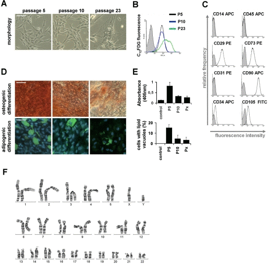
Figure 2.Changes of MSC during culture expansionSenescent MSC acquire a typical large and flat morphology, whereas no differences were observed at passage 5 and passage 10 (A). Expression of the senescence-associated β-galactosidase was detected with the fluorogenic substrate C12FDG and this biomarker for replicative senescence was positive in senescent passages but not in passage 5 and passage 10 (B). All MSC preparations displayed the typical immunophenotype (C) and could be induced towards osteogenic and adipogenic lineage (D). However, this in vitro differentiation potential decayed already between passage 5 and passage 10 (E; control is without induction medium; Px represents the corresponding senescent passage; all data are presented as mean ± SD; n = 4). A normal karyogram of MSC (after treatment with trypsin and Giemsa)is exemplarily presented (F). (scale bars = 100μm).