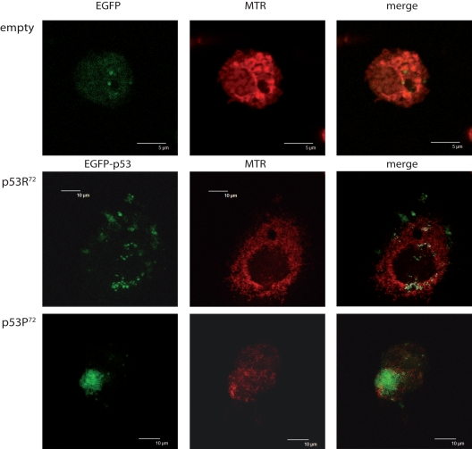
Figure 1.Different localisation of p53 isoforms after rotenone treatment. p53−/− HCT116 cells transfected with empty EGFP pCMS plasmid (upper panels), EGFP-p53R72 pCMS plasmid (central panels), or EGFP-p53P72 pCMS plasmid (lower panels) and counterstained with MitoTracker Red (MTR). Cells transfected with the empty vector show a diffused EGFP fluorescence, not associated with any cell structure; in cells transfected with the plasmids expressing the p53 isoforms, a different subcellular localisation is noticed, as assessed by counter-staining with the mitochondrial-specific probe MTR. In particular, when EGFP-p53R72 pCMS plasmid is used, EGFP fluorescence, associated to p53 protein, appears to be not nuclear but rather co-localised with MTR fluorescence, indicating that p53 has a mitochondrial localisation (white dots indicate points of overlapping of the two fluorescences).