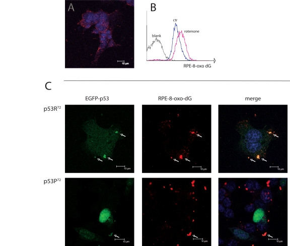
Figure 2.Co-localisation of p53 isoforms with damaged mtDNA. (A) p53−/− HCT116 cells treated with 100 nM rotenone for 24 hours and stained for 8-oxo-dG and revealed with RPE-conjugated secondary moAb (red fluorescence). Nuclei are counterstained with Hoechst 33258. Punctuated, cytoplasmic red fluorescence indicates that 8-oxo-dG accumulates in mitochondria but not nuclei upon rotenone treatment. (B) flow cytometric detection of 8-oxo-dG after rotenone treatment. C: p53−/− HCT116 cells transfected with either EGFP-p53R72 or EGFP-p53P72 pCMS plasmid and treated as in A. Arrows indicate the points in which 8-oxo-dG (red fluorescence) and p53 (green fluorescence) co-localise (yellow dots in the merged picture). Note that in the case of p53R72 the green fluorescence is diffused in the cytoplasm and tends to accumulate in mitochondria, while in the case of p53P72 the green fluorescence is mainly localised to the nucleus.