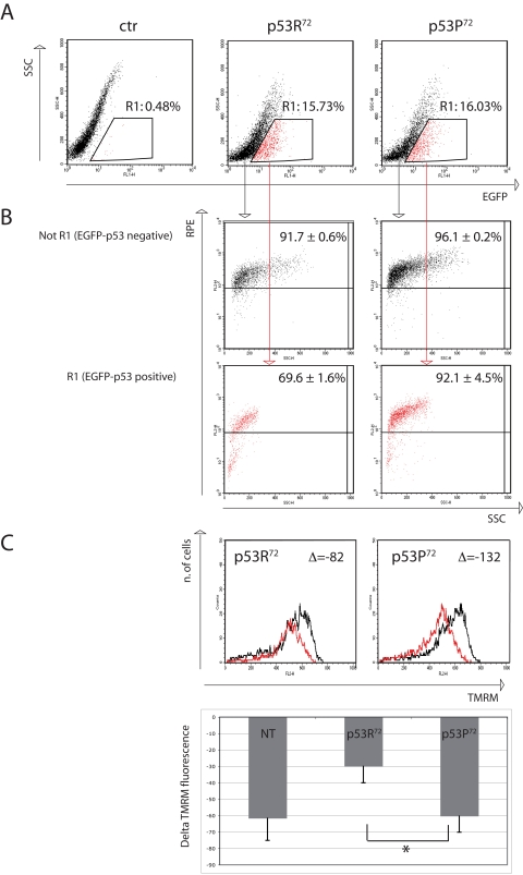
Figure 3.Flow cytometry analysis of p53−/− HCT116 cells transfected with either EGFP-p53R72 or EGFP-p53P72 pCMS plasmid. After 24 hours from transfection, cells were treated with 100 nM rotenone and incubated for additional 24 hours, then stained for 8-oxo-dG or TMRM (see materials and methods). (A) transfection efficiency. Cells contained in R1 are considered EGFP-positive. Ctr: non-transfected cells. (B) 8-oxo-dG detection. The cells in (R1) and (not R1) are evaluated for 8-oxo-dG fluorescence (RPE). Numbers represent the percentage of cells with high 8-oxo-dG fluorescence and are expressed as mean ± st. dev. of three independent experiments. As showed EGFP-negative cells (not R1, not expressing p53) are almost all positive for 8-oxo-dG fluorescence (more than 90%), while when considering EGFP-positive cells (R1, expressing p53), the cells that were transfected with p53R72 which resulted positive for 8-oxo-dG decreased to 69%, with respect to 92% of those that were transfected with p53P72. See text for comment. (C) Mitochondrial membrane potential (MMP) analysis. The cells in (R1) were evaluated for MMP by using the potentiometric dye TMRM. Black line: control cells; red line: rotenone treatment. Decrease in MMP was expressed as the difference (Δ) of TMRM fluorescence intensity between rotenone-treated and control cells. Cells positive for EGFP-p53R72 (left panel) display a lower decrease in MMP with respect to those positive for EGFP-p53P72 (right panel). The graphic shows data related to 3 independent experiments (mean ± st. dev.) * Student' t test p= 0.022. NT, not transfected.