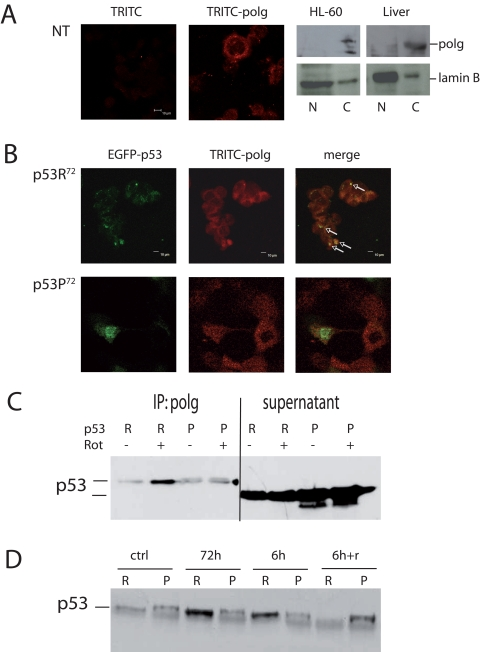
Figure 4.Co-localisation of p53 and polg. (A) Confocal analysis of p53−/− HCT116 non-transfected cells stained with anti-polg antibody alone or with the TRITC-conjugated secondary antibody; Western Blot analysis of specificity of the anti-polg antibody on cytoplasmic and nuclear fractions obtained from HL-60 cells and human hepatocytes. As shown, the band corresponding to the molecular weight of polg is present only in the cytoplasmic fractions. (B) Confocal analysis of p53−/− HCT116 cells transfected with EGFP-p53R72 (upper panels) or EGFP-p53P72 (lower panels) pCMS plasmids and treated with 100 nM rotenone for 24h. It is possible to observe that only in the case of p53R72 many points of co-localisation are visible (arrows). (C, D) Co-Immunoprecipitation assay on stably transfected PC3 cells. In C cells were treated for 72h with 10 nM rotenone. R= p53R72; P= p53P72. The lanes indicated as supernatant show the amount of p53 expressed by the cells. In D cells were exposed to 10 nM rotenone for 6h, 72h, 6h and recovery until 72h (6h+r).