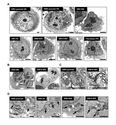
Figure 4.Morphological variations of patient-derived iPS cells upon reprogramming(A) High-resolution electron micrographs of HK cells before (SW4 parental HK and SW8 parental HK) and after (SW4 #N1, SW3 #B, SW8 #20I and SW10 #5P) induced pluripotency. Representative micrograph of a verified fibroblast-derived iPS cell is also included. Scale bars represent 2 μm. (B) Mitotic events of two iPS clones were shown (left panel in metaphase; right panel in anaphase). Scale bars represent 2 μm. (C) Endoplasmic reticulum and the Golgi structures in HK and HK-derived iPS cells are shown. Scale bars represent 0.5 μm. (D) Mature mitochondria with well-developed cristae in parental HK cells (SW8 parental) and immature mitochondria in iPS clones (SW3 #B, SW8 #20I and SW10 #5P) are indicated by arrows. Keratin intermediate filaments in parental HK cells are indicated by arrowheads. Scale bars represent 0.5 μm.