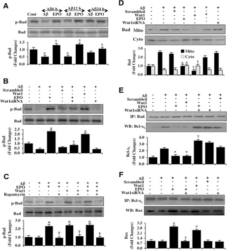
Figure 4.EPO through Wnt1 phosphorylates Bad, controls mitochondrial trafficking of Bad, and modulates Bad, Bcl-xL, and Bax binding(A) Microglial protein extracts (50 μg/lane) were immunoblotted with phospho-rylated Bad (p-Bad, (Ser136)) antibody at 6, 12, and 24 hours following administration of Aβ (10 μM). Aβ exposure resulted in a significant decrease in the expression of p-Bad. EPO (10 ng/ml) with a 1 hour pretreatment significantly increased the expression of p-Bad 6 hours following Aβ exposure (*P <0.01 vs. Control; †P<0.01 vs. Aβ of corresponding exposure time). In all cases, each data point represents the mean and SEM from 3 experiments. (B) Gene reduction of Wnt1 was performed with transfection of Wnt1 siRNA prior to Aβ (10 μM) administration in microglia. The expression of p-Bad was determined at 6 hours following Aβ exposure. EPO (10 ng/ml) or Wnt1 (100 ng/ml) with 1 hour pretreatments significantly increased the expression of p-Bad 6 hours following Aβ exposure. Yet, Wnt1 siRNA transfection prior to Aβ exposure prevented EPO from significantly phosphorylating Bad. Non-specific scrambled siRNA did not significantly alter p-Bad expression during Aβ exposure (*P < 0.01 vs. Aβ; †P<0.01 vs. EPO/Aβ). (C) EPO (10 ng/ml), Wnt1 (100 ng/ml) or combined EPO with Wnt1 with 1 hour pretreatments significantly increased the expression of p-Bad 6 hours following Aβ (10 μM) exposure. Application of the mTOR inhibitor rapamycin (50 nM) with EPO (10 ng/ml), Wnt1 (100 ng/ml) or combined EPO with Wnt1 1.5 hour prior to Aβ exposure resulted in the loss of the ability of EPO, Wnt1, or combined EPO with Wnt1 to increase in the expression of p-Bad during Aβ exposure (*P < 0.01 vs. Aβ; †P<0.01 vs. EPO/Aβ, Wnt1/Aβ or EPO/Wnt1/Aβ). (D) Gene reduction of Wnt1 was performed with transfection of Wnt1 siRNA prior to Aβ (10 μM) administration in microglia and the expression of Bad in both cytosolic and mitochondrial fractions was determined at 6 hours following Aβ exposure. EPO (10 ng/ml) or Wnt1 (100 ng/ml) with 1 hour pretreatments significantly reduced mitochondrial expression of Bad and increased the cytosolic expression of Bad following Aβ exposure. Yet, gene reduction of Wnt1 with Wnt1 siRNA transfection led to the loss of the ability of EPO to promote the release of Bad for the mitochondria to the cytosol during Aβ exposure. Non-specific scrambled siRNA did not significantly change the translocation of Bad during Aβ exposure (*P < 0.01 vs. Aβ; †P < 0.01 vs. EPO/Aβ). (E) Gene reduction of Wnt1 was performed in microglia with transfection of Wnt1 siRNA prior to Aβ (10 μM) administration. Protein extracts were immunoprecipitated using Bad antibody 6 hours following Aβ exposure. Western blot for Bcl-xL expression in the precipitates was performed. EPO (10 ng/ml) or Wnt1 (100 ng/ml) with 1 hour pretreatments decreased binding of Bcl-xL to Bad during Aβ exposure. Gene reduction of Wnt1 with Wnt1 siRNA transfection prevented EPO to decrease the binding of Bcl-xL to Bad during Aβ exposure. Non-specific scrambled siRNA did not significantly alter the binding of Bad to Bcl-xL during Aβ exposure (*P < 0.01 vs. Aβ; †P<0.01 vs. EPO/Aβ). (F) Gene reduction of Wnt1 was performed with transfection of Wnt1 siRNA prior to Aβ (10 μM) administration in microglia. Protein extracts were immunoprecipitated using Bcl-xL antibody 6 hours following Aβ exposure. Western blot for Bax expression in the precipitates was performed. EPO (10 ng/ml) or Wnt1 (100 ng/ml) with 1 hour pretreatments increased the binding of Bcl-xL to Bax during Aβ exposure. Gene reduction of Wnt1 with Wnt1 siRNA transfection prevented EPO to increase the binding of Bcl-xL to Bax during Aβ exposure. Non-specific scrambled siRNA did not significantly alter the binding of Bcl-xL with Bax during Aβ exposure (*P < 0.01 vs. Aβ; †P<0.01 vs. EPO/Aβ).