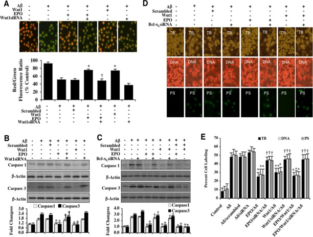
Figure 5.EPO and Wnt1 control mitochondrial membrane potential, block early and late apoptotic microglial Aβdegeneration, and prevent caspase 1 and 3 activation through Bcl-xL(A) Representative images and quantitative results from JC-1 staining illustrate that Aβ (10 μM)results in a significant decrease in the red/green fluorescence intensity ratio of mitochondria within 6 hours when compared with untreated control cultures, demonstrating that Aβ exposure leads to significant mitochondrial membrane depolarization. EPO (10 ng/ml) or Wnt1 (100 ng/ml) with 1 hour pretreatments significantly increase the red/green fluorescence intensity of mitochondria in microglia, demonstrating that mitochondrial membrane potential was restored. In contrast, gene reduction of Wnt1 with transfection of Wnt1 siRNA increased mitochondrial membrane depolarization to a greater degree than Aβ exposure alone and prevented the ability of EPO to maintain mitochondrial membrane potential during Aβ exposure. The relative ratio of red/green fluorescent intensity of mitochondrial staining was measured in 6 independent experiments with analysis performed using the public domain NIH Image program (http://rsb.info.nih.gov/nih-image) (*P<0.01 vs. Aβ; †P <0.01 vs. EPO/Aβ). (B) Microglial cell protein extracts (50 μg/lane) were immunoblotted with cleaved caspase 1 (active) and cleaved caspase 3 (active) antibodies 6 hours following Aβ(10 μM) exposure.Aβ(10 μM) exposure significantly increased caspase 1 and caspase 3 activities. In contrast, EPO (10 ng/ml) or Wnt1 (100 ng/ml) administration significantly decreased the expression of cleaved (active) caspase 1 and caspase 3 at 6 hours following Aβ(10 μM) exposure. Gene reduction of Wnt1 with transfection with Wnt1 siRNA abrogated the ability of EPO to prevent caspase activation (*P<0.01 vs. Aβ; †P <0.01 vs. EPO/Aβ). Non-specific scrambled siRNA did not significantly change the expression of cleaved caspase 1 and caspase 3 during Aβ exposure. Each data point represents the mean and SEM from 3 experiments. Quantification of western band intensity from 3 experiments was performed using the public domain NIH Image program (http://rsb.info.nih.gov/nih-image). (C) Microglial cell protein extracts (50 μg/lane) were immunoblotted with cleaved caspase 1 (active) and cleaved caspase 3 (active) antibodies 6 hours following Aβ(10 μM) exposure.Aβ(10 μM) exposure significantly increased caspase 1 and caspase 3 activities. In contrast, EPO (10 ng/ml) or Wnt1 (100 ng/ml) administration significantly decreased the expression of cleaved (active) caspase 1 and caspase 3 at 6 hours following Aβ(10 μM) exposure. Gene reduction of Bcl-xLwith transfection with Bcl-xL siRNA abrogated the ability of EPO to prevent caspase activation (*P<0.01 vs. Aβ; †P <0.01 vs. EPO/Aβ). Non-specific scrambled siRNA did not significantly change the expression of cleaved caspase 1 and caspase 3 during Aβ exposure. Each data point represents the mean and SEM from 3 experiments. Quantification of western band intensity from 6 experiments was performed using the public domain NIH Image program (http://rsb.info.nih.gov/nih-image). (D and E)EPO (10 ng/ml) or Wnt1 (100 ng/ml) were administered to microglial cultures 1 hour prior to the Aβ (10 μM) exposure and trypan blue dye exclusion, DNA fragmentation, and membrane PS exposure were determined 24 hours later.Representative images (B) and quantitative analysis (C) demonstrate that Aβ(10 μM) results in a significant increase in trypan blue staining, DNA fragmentation, and membrane PS exposure in microglia 24 hours after Aβ exposure compared to untreated control cultures. In contrast, EPO (10 ng/ml), Wnt1 (100 ng/ml), or combined EPO and Wnt1 applied 1 hour prior to Aβ significantly reducedtrypan blue staining, DNA fragmentation, and membrane PS exposure in microglia 24 hours after Aβ exposure. Gene reduction of Bcl-xLwith transfection of Bcl-xL siRNA prior to Aβ exposure prevented EPO (10 ng/ml), Wnt1 (100 ng/ml), or combined EPO and Wnt1 from blocking cell injury and resulted in increased trypan blue staining, DNA fragmentation, and membrane PS exposure in microglia 24 hours following Aβ exposure. Non-specific scrambled siRNA did not significantly change cell injury during Aβ exposure (*P < 0.01 vs. Aβ; †P <0.01 vs. EPO/Aβ, Wnt1/Aβ, or EPO/Wnt1/Aβ). Each data point represents the mean and SEM from 6 experiments.