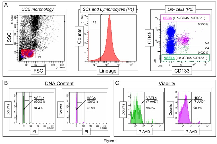
Figure 1.Morphological characteristics of UCB-derived VSELs by flow cytometric assaysPanel A shows a well-established flow cytometric protocol for identification of CD133+Lin−CD45− VSELs among the whole-nucleated fraction of UCB cells. The dot-plot (left) visualizes cells based on FSC and SSC parameters, indicating the relative cellular size and complexity, respectively. The FSClow/SSClow lymphocytic population, which includes very small objects, is enclosed in region P1, and the cells were further analyzed according to hematopoietic lineage marker expression (middle histogram). Lin- cells from region P2 are plotted in the dot-plot representing CD133 and CD45 expression (right). VSELs are identified as CD133+Lin−CD45− cells (region Q4), while HSCs are identified as CD133+Lin-CD45+ cells (region Q2). Percentages indicate the content of both stem cell populations among all nucleated cells in one representative UCB sample. Panel B presents a representative analysis of DNA content in UCB-derived VSELs and HSCs following fixation and staining with propidium iodide (PI). The percentages indicate normal diploid (2n) VSEL and HSC fractions in the G0/G1 phase of the cell cycle. Panel C shows the viability of VSELs and HSCs examined by flow cytometry following the staining of freshly isolated cells with a viability dye, 7-aminoactinomycin D (7-AAD). Viable VSELs and HSCs are represented as 7-AAD− cells, which exclude the dye, and their percentage content is computed for each of the stem cell populations.