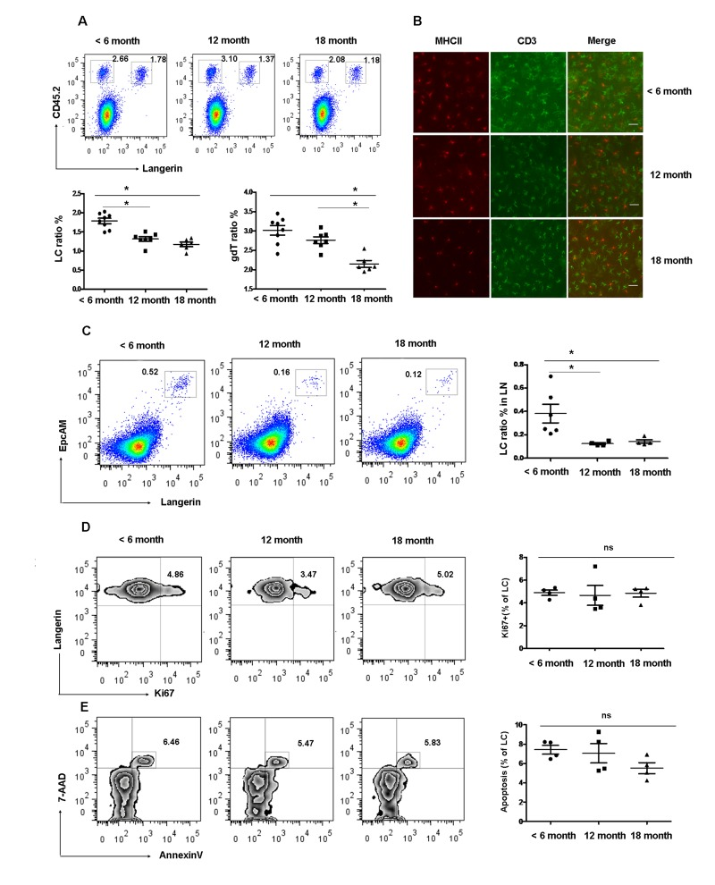
Figure 1.Gradual loss of epidermal LCs during aging development(A) and (B) Reduced epidermal LCs and DETCs during aging development. The number of Langerin+CD45.2+epidermal LCs and Langerin−CD45.2+ DETCs were analyzed by FACS (A). Data are shown as mean±SEM of 6-8 mice at each time point. MHCII+ LCs and CD3+ DETCs in the epidermis was assessed by imunofluorescence at each time point (B). Scale bar, 10μm, original magnification (200×). (C) Migrated LCs in draining LNs. Cells from LNs were stained by Anti-IAE, CD8, EpCAM, and Langerin. The EpCAM+Langerin+ migrated LCs were analyzed on gated CD8−IAE+ cells. Data are also shown as mean±SEMof 4-6 mice at each time point. *p < 0.05. (D) The kinetics of appearance of Ki67-labeled LCs in aged and young mice was determined by FACS analysis. (E) Apoptotic epidermal LCs were elucidated using Annexin-V antibody and the viable stain 7-AAD. Data are shown as mean ± SEM of 4 mice at each time point and representative of 2 independent experiments.