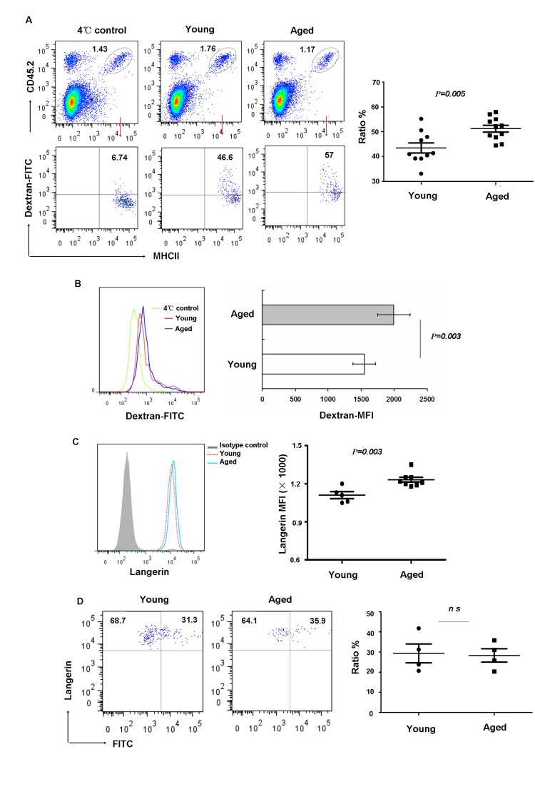
Figure 3.Aging impaired LC phagocytic ability but did not affect migration in vivo(A-C) Epidermal cells were incubated with 0.25 mg/ml FITC-Dextran for 45min at 4°C (control) or 37°C, and then stained with anti-MHCII and anti-CD45.2 antibodies. The percentage of FITC+ cells in LCs (CD45.2+MHCII+ cells) was determined (A). The geometric mean fluorescence of FITC expression in LCs was also shown (B) and the expression of Langerin in aging LCs was shown (C).(D) Aged LC migration. Mice were painted with 200μl 5mg/ml FITC in acetone/dibutylphthalate on the abdomen. LN cells collected 24hrs later were stained with anti-CD8, anti-Langerin and anti-EpCAM antibodies. The ratio of migrated FITC+ LCs was analyzed on gated Langerin+EpCAM+ LCs. Data are representative of 2 independent experiments, 4 mice were analyzed in each experiment. ns represents non-statistical significance.