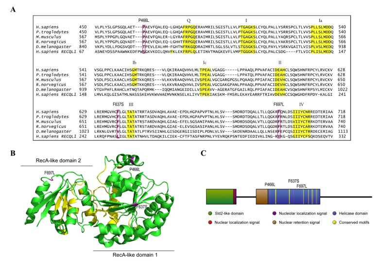
Figure 1.Homology map and model structure of RECQL4(A) RECQL4 homologues across several species, illustrating the highly conserved nature of the amino acids examined in this study. Important helicase motifs are highlighted in yellow [50], while mutations are boxed in magenta. The sequence of RECQL1 is included to show the alignment used to create the model structure in B. (B) The crystal structure of RECQL1 without the RQC domain that is not present in RECQL4 (PDB ID: 2WWY), and with the amino acids homologous to the examined mutations in RECQL4 highlighted in magenta. Labels indicate the mutations in RECQL4. Important helicase motifs are highlighted in yellow, and the RecA-like domains 1 and 2 are indicated. (C) Domain map of RECQL4, with the examined mutations highlighted in magenta, and with important helicase motifs highlighted in yellow.