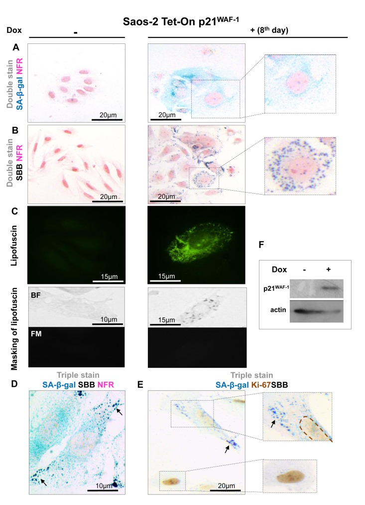
Figure 2.Lipofuscin accumulates and co-localizes with Senescence-Associated beta-galactosidase (SA-β-gal) in senescent Saos-2 cells triggered by p21WAF-1(A) SA-β-gal staining (turquoise color) in the Saos-2 p21WAF-1 Tet-On cell system on the 8th day of doxycycline (5 μg/ml) addition. Inset: Senescent cells acquired the characteristic senescent morphological phenotype (enlarged and flattened). (B) Sudan Black B (SBB) positivity (dark blue-black granules) in cells with senescent morphological phenotype (inset). (C) Top panels: Lipofuscin's auto-fluorescence in induced Saos-2 p21WAF-1 Tet-On cells, by fluorescence microscopy at 450-490 nm (green pseudocolor). Bottom panels: Cytochemical SBB staining (BF, bright field microscopy) quenches the auto-fluorescence of lipofuscin (FM, fluorescence microscopy), indicating that SBB stains lipofuscin. SA-β-gal and SBB staining coincided in cells that had the morphological phenotype of senescence (D) and were absent in cells that were positive for the proliferative marker Ki67 (E). (F) Addition of doxycyclin (Dox) triggers p21WAF-1 expression. Brown dashed lines: Ki67- negative nuclei. Black arrows: SBB granules. NFR: nuclear fast red counterstain.