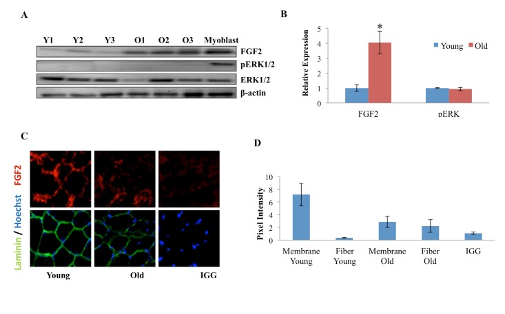
Figure 3.Age-dependent comparison of FGF2 and pERK levels and localization in muscle fibers(A) Protein was isolated from freshly-derived uninjured myofibers of young and old mice and the levels of FGF2 and phospho-ERK1/2; total ERK1/2 and cytoplasmic beta-actin were analyzed by Western blotting, using specific antibodies. Representative data are shown. (B) Relative protein expression was quantified in 3 young and 3 old mice by normalization of FGF-2 to beta-actin and normalization of pERK to total ERK; significantly higher levels of FGF-2, but not of pERK were detected in the old myofibers, as compared to young (n=3, * P<0.05). (C) Tibialis anterior (TA) muscle from 2 young and 2 old mice were sectioned and immunostained for laminin (green) and FGF2 (red). Hoechst (blue) labels all nuclei. Representative images demonstrate the presence of FGF-2 and laminin in muscle compartments, as compared to the negative IgG control and higher FGF-2 levels seem to be present in the laminin+ basement membranes of the young myofibers, as compared to old. (D). The pixel density of FGF-2 that co-localizes with laminin+ basement membrane vs. the internal regions of the myofibers was determined in 30-40 areas of each cryosection of 3 muscle tissue slides from young and old muscle, using Image J software. Preferential localization of FGF-2 in the basement membrane was identified in young muscle, while in the old tissue, FGF-2 was mis-localized to the center of the myofibers and away from the basement membrane, n=3, * P<0.05.