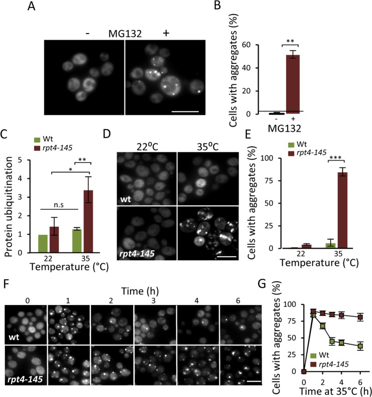
Figure 2.Lowering proteasome function results in increased protein aggregation.(A) Representative images of Hsp104-GFP distribution with and without the addition of the proteasome inhibitor MG132 (100 μM) to a growing culture. (B) Percentage of cells with Hsp104-GFP foci after partial proteasomal inhibition by MG132. (C) Relative levels of protein ubiquitination upon growing the conditional proteasomal mutant rpt4-145 (ts) at the permissive (22°C) and near non-permissive (35°C) temperature. Levels were normalized to the levels in wt cells grown at 22°C. (D) Representative images of Hsp104-GFP localization upon growth of wt and rpt4-145 cell at the permissive (22°C) and near non-permissive (35°C) temperature. (E) Percentage of wt and rpt4-145 cells with Hsp104-GFP foci after growth at the permissive (22°C) and near non-permissive (35°C) temperature. (F) The clearance of Hsp104-GFP foci was followed over time after an initial burst in aggregate formation after the temperature shift. Time point “0” depicts cells growing at 22°C and subsequent time points depict cells following the indicated time at 35°C. (G) Percentage of wt and rpt4-145 cells with Hsp104-GFP foci. Quantification of Hsp104-GFP foci formation in the experiment in “F”. Error bars represent standard deviation (n=2). For statistical analysis, the paired two-tailed t-test was used where *P<0.05, **P<0.01, ***P<0.001 and n.s = no significant difference. (n= sets of analysis; Scale-bars represent 10μm).