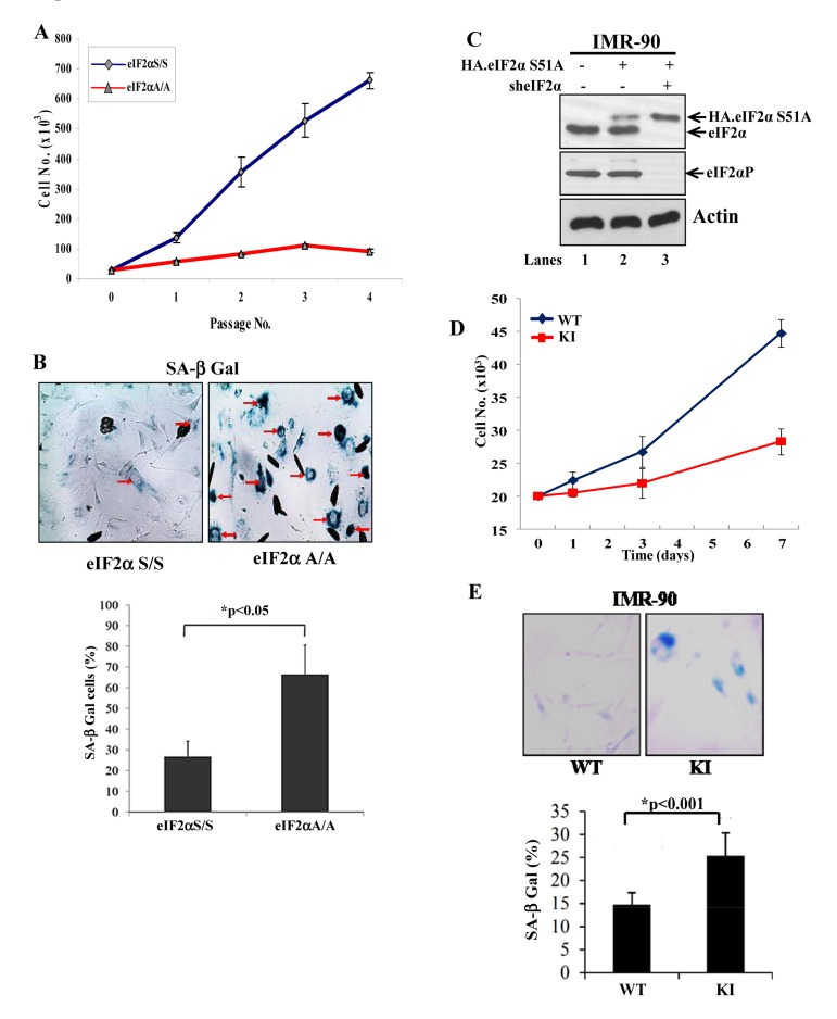
Figure 1.Loss of eIF2αP impairs proliferation and induces premature senescence in primary cells(A) Primary eIF2αS/S and eIF2αA/A MEFs were maintained in culture for the indicated passages. Proliferation was measured by cell counting. Values represent an average taken from two independent experiments performed in triplicates. (B) Senescence in eIF2αS/S and eIF2αA/A MEFs was monitored by SA β-Gal staining. The ratio of the percentage of SA-β-gal positive cells obtained from two independent experiments is as indicated in the histogram. Senescent cells are indicated by arrows. (C) Primary IMR90 human fibroblasts were engineered to express an HA-tagged eIF2αS51A (lanes 2 and 3) under conditions in which endogenous eIF2α was down regulated by shRNA expression (lane 3; KI cells). Protein extracts (50 μg) were subjected to immunoblot analysis for the indicated proteins. Note that the slow-migrating band detected by the anti-eIF2α antibody corresponds to HA-eIF2αS51A (lanes 2, 3) and that endogenous eIF2α was substantially down regulated by shRNA expression (lane 3). Control IMR90 cells (WT) in lane 1 represent cells which were infected with insert-less retroviruses and lentiviruses. (D) Proliferation of IMR90 WT and KI cells was measured by cell counting. Values represent an average of two independent experiments performed in triplicates. (E) Cells were stained for SA β-Gal and the average percentage values of positive cells from three independent experiments are indicated. Senescent cells are in blue color.