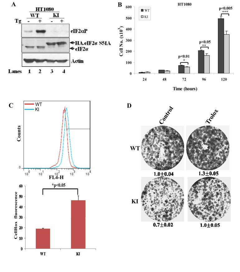
Figure 6.Inactivation of eIF2αP in human tumor cells decreases cell proliferation and increases ROS levels(A) Cell extracts (50 μg of protein) from wild-type (WT) and knock-in (KI) HT1080 cells treated with 1μM thapsigargin (TG) for 2 hours were subjected to immunoblot analysis for the indicated proteins. Note the slower migration of the HA-eIF2αS51A in KI cells (lanes 3,4) compared to endogenous eIF2α in WT cells (lanes 1,2). (B) Cell proliferation of HT1080 WT and KI cells was assessed by cell counting and was plotted over time. The error bars indicate the standard deviation. (C) ROS levels in HT1080 WT and KI cells was assessed by Cell-RoxTM Deep Red staining and FACS analysis. Histograms represent the average ROS levels measured by CellRox fluorescence from three independent experiments. (D) Colony formation assays of HT1080 WT and KI cells in the absence or presence of 200 μM Trolox. Cells were stained with crystal violet. Values represent ratios of optical density (OD) in arbitrary units.