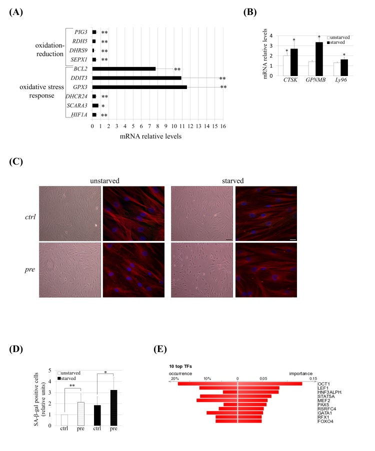
Figure 4.hMSCs show an altered transcriptomic profile and phenotype of senescence under prelamin A accumulation and serum starvation conditions. (A) Q-RT-PCR validation for a subset of genes grouped in oxidation-reduction and response to oxidative stress categories and (B) for genes known to be up-regulated in senescent hMSCs. The gene expression ratio for pre-hMSCs versus control-hMSCs under starved conditions is shown in A and in the case of B it has been included too the comparison of pre-hMSCs versus ctrl-hMSCs ratio under unstarved conditions. For gene expression normalization GAPDH was used. Bars are mean +/- standard error mean of 3 independent donors. ** p< 0.01, * p<0.05. (C) Senescence-associated morphological changes in pre-hMSCs under serum starvation conditions. Bright field images (scale bar: 100 μm) and confocal immunofluorescence images (scale bar: 20 μm) are shown. Red: beta-tubulin, blue: DAPI. (D) SA β-gal quantification in hMSCs under basal (unstarved) or serum starvation conditions (starved). Bars are average +/- standard deviation. ** p< 0.01, * p<0.05. (E) DiRE analysis of genes found to be dys-regulated in pre-hMSCs. The graph shows the top 10 candidate transcription factors ranked by importance.
(pre): prelamin A-accumulating hMSCs, (ctrl): control-hMSCs.