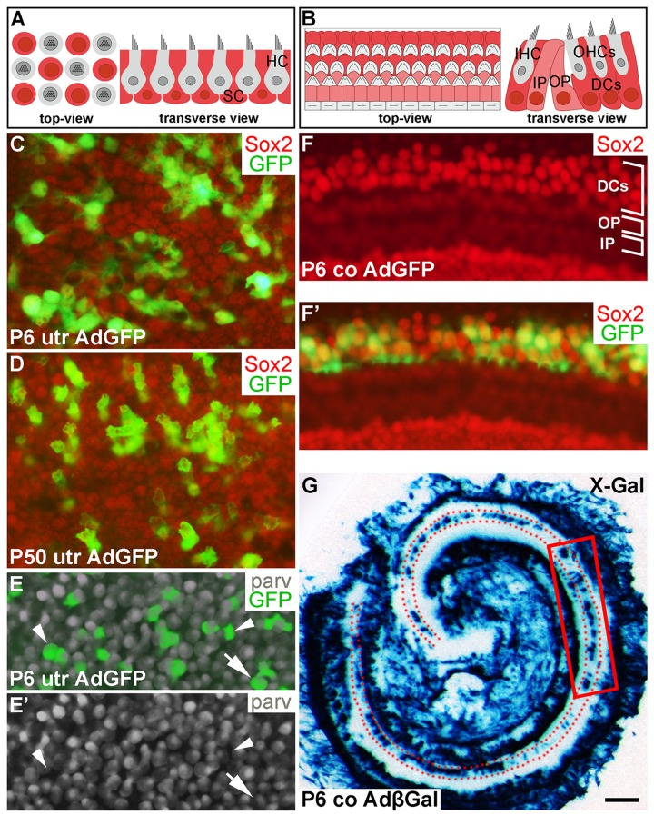
Figure 1.Adenoviruses transduce inner ear supporting cells in explant cultures. AdGFP- and AdβGal-infected utricles and cochleas analyzed after 3 DIV. (A,B) Schematic representation of the utricular (A) and cochlear (B) sensory epithelium, viewed from above (whole mount specimens) and in transverse plane. Utricular hair cells with the apical stereociliary bundle (grey) are located on top of a layer SCs (red). The cochlear sensory epithelium consists of one row of inner hair cells and three rows of outer hair cells (grey). Deiters' cells (red) are located underneath outer hair cells. Inner and outer pillar cells (pink) are positioned between the inner and outer hair cell rows. (C,D) AdGFP-infected P6 and P50 utricles double-labeled for GFP and Sox2 show transduction in SCs. The views are focused to the level of Sox2+ SC nuclei. (E,E') In AdGFP-infected P6 utricle, a small part of parvalbumin+ hair cells are transduced (arrow), in addition to SCs (arrowheads). (F,F') In P6 cochlea, Deiters' cells show AdGFP transduction, as opposed to the adjacent outer and inner pillar cells. (G) X-Gal histochemical staining shows a patchy pattern of AdβGal transduction in the area of Deiters' cells (dotted) along the length of the cochlear duct. The boxed area represents the region used for analysis. Abbreviations: utr, utricle; co, cochlea; AdβGal, adenovirus encoding β-galactosidase; AdGFP, adenovirus encoding green fluorescent protein; parv, parvalbumin; DCs, Deiters' cells; IP, inner pillar cell; OP, outer pillar cell; IHC, inner hair cell; OHCs, outer hair cells. Scale bar, shown in G: C-F', 20 µm; G, 180 µm.